About LJVascular

This author has not yet filled in any details.
So far LJVascular has created 514 blog entries.

21, 12, 2023

Unveiling the Road to Better Health: The Power of Carotid Artery Ultrasound

2023-10-11T17:22:03-07:00

Unveiling the Road to Better Health: The Power of Carotid Artery Ultrasound

Envision having the ability to peer into the intricate passageways that serve as vital conduits for blood traveling to your brain, allowing you to detect potential threats before they strike. Carotid Artery Ultrasounds provide precisely this capability, offering a non-invasive window into your carotid arteries to fortify the defense against strokes. In this blog post, we shall embark on a journey into the realm of carotid artery ultrasounds, elucidating their critical role in stroke prevention and acquainting you with the risk factors that demand your attention.

Unveiling the Significance of Carotid Artery Ultrasounds

Carotid Artery Ultrasounds are more than mere routine assessments; they represent powerful weapons in the battle against strokes. These ultrasounds serve as vital instruments for appraising the condition of the carotid arteries, responsible for supplying the brain with oxygen-rich blood. Their primary mission is to unearth potential issues that may pave the way for strokes, particularly in individuals who have manifested symptoms of a Transient Ischemic Attack (TIA) or are perched atop the precipice of stroke risk.

Deciphering the Transient Ischemic Attack (TIA)

Before we immerse ourselves in the intricacies of carotid artery ultrasounds, it’s essential to shed light on Transient Ischemic Attacks, commonly known as TIAs. These episodes serve as warnings that the brain’s blood supply faces a momentary interruption. The symptoms associated with TIAs are akin to those of a stroke and may encompass:

  • Sudden numbness in the face, arm, or leg, especially on one side of the body.
  • Difficulty speaking or slurred speech.
  • Vision anomalies, such as abrupt blurred or double vision.
  • Dizziness or loss of balance.
  • An intense headache without any apparent cause.

TIAs mirror the symptoms of a stroke, but the fundamental distinction lies in their transient nature. TIAs are ephemeral episodes, typically lasting only a few minutes to a couple of hours, and they do not inflict permanent damage. However, they act as substantial red flags, signaling the potential for a forthcoming stroke.

Assessing Stroke Risk through Ultrasound Screenings

Carotid artery ultrasounds prove instrumental in the assessment of stroke risk, particularly in individuals who have encountered TIA symptoms. Here is how these screenings operate:

  • Non-Invasive Imaging: Carotid artery ultrasounds harness non-invasive imaging technology to render the carotid arteries in the neck visible. By using high-frequency sound waves, these ultrasounds craft detailed images of these pivotal blood vessels.
  • Discerning Blockages: The ultrasound aids in the detection of blockages or the buildup of plaque within the carotid arteries. These impediments can diminish the blood flow to the brain, amplifying the vulnerability to a stroke.
  • Early Intervention: The early detection of blockages empowers healthcare providers to take preemptive actions aimed at stroke prevention. This may encompass the administration of medications or surgical procedures designed to clear the arteries.

Familiarize Yourself with the Risk Factors for Strokes and Arterial Disease

Gaining a comprehensive understanding of the risk factors associated with strokes and arterial disease represents the initial stride on the path to prevention. The following risk factors warrant your attention:

  • High Blood Pressure: Hypertension reigns as a principal contributor to stroke, underscoring the imperative nature of blood pressure management.
  • Tobacco Usage: Smoking escalates the risk of arterial disease and strokes considerably. The act of quitting smoking ranks among the most effective measures for curbing this risk.
  • Diabetes: Individuals contending with diabetes encounter an elevated risk of strokes. Prudent management of blood sugar levels assumes paramount significance.
  • Family History: A familial history intertwined with strokes or arterial disease can elevate your risk.
  • Age: The risk of a stroke surges with age, particularly following the age of 55.
  • Obesity: Excessive weight may contribute to heightened blood pressure and various other risk factors.
  • Sleep Apnea: Untreated sleep apnea can elevate the risk of stroke due to interrupted breathing during sleep.

Carotid Artery Ultrasounds transcend their identity as mere medical procedures; they stand as vigilant protectors of your cerebral well-being. If you have encountered TIA symptoms or belong to the categories at increased risk, the discussion of carotid artery ultrasounds with your healthcare provider becomes a proactive step toward stroke prevention. Through the comprehension of warning signs and risk factors, you retain the power to command your vascular health and reduce the likelihood of a stroke rewriting the narrative of your life.

“Bringing Experts Together for Unparalleled Vein and Vascular Care”

La Jolla Vein & Vascular (formerly La Jolla Vein Care) is committed to bringing experts together for unparalleled vein and vascular care. 

Nisha Bunke, MD, Sarah Lucas, MD, and Amanda Steinberger, MD are specialists who combine their experience and expertise to offer world-class vascular care. 

Our accredited center is also a nationally known teaching site and center of excellence. 

For more information on treatments and to book a consultation, please give our office a call at 858-550-0330. 

 For a deeper dive into vein and vascular care, please check out our Youtube Channel at this link, and our website https://ljvascular.com

For more information on varicose veins and eliminating underlying venous insufficiency,

Please follow our social media Instagram Profile and Tik Tok Profile for more fun videos and educational information. 

For more blogs and educational content, please check out our clinic’s blog posts

Unveiling the Road to Better Health: The Power of Carotid Artery Ultrasound2023-10-11T17:22:03-07:00

Guardians of Life: Nurturing Arterial Health for a Vibrant Tomorrow

2023-10-11T17:12:31-07:00

Guardians of Life: Nurturing Arterial Health for a Vibrant Tomorrow

Within our intricate vascular system, arteries act as the vital highways, ensuring the efficient transportation of oxygen-rich blood throughout our bodies. This network plays a pivotal role in sustaining the proper functioning of our organs and tissues. However, the passage of time may present challenges to these indispensable thoroughfares, manifesting as issues like plaque buildup and blockages. In this blog post, we shall embark on a journey to understand the importance of arterial testing and screening, the diverse diagnostic methods available, and those individuals who should consider these crucial examinations.

Exploring Arterial Testing and Screening

Arterial testing and screening stand as foundational elements in the realm of vascular health assessment. These examinations furnish us with invaluable insights into the state of our arteries and serve as early detectors for concerns such as plaque accumulation, blockages, and reduced blood flow. Let us delve into some of the key diagnostic methods at our disposal:

  • Ankle-Brachial Index (ABI): ABI is a non-invasive evaluation that scrutinizes blood flow in both the legs and arms, employing inflatable pressure cuffs. This method yields vital information about the presence of arterial blockages and contributes to an overall assessment of arterial health.
  • Duplex Ultrasound: Duplex ultrasound emerges as a versatile instrument for scrutinizing the flow of blood through arteries and veins. This imaging tool encompasses various ultrasound tests, tailored to specific areas of interest:
    • Lower Extremity Arterial Study (LEA): This ultrasound zooms in on the arteries located in the lower legs, aiming to detect plaque buildup and potential blockages, often linked to atherosclerosis.
    • Upper Extremity Arterial Study (UEA): UEA takes a closer look at the arteries residing in the upper arms and neck, delivering insights into the state of arterial health in these regions.
    • Carotid Artery Ultrasound: This ultrasound focuses on assessing the carotid arteries in the neck, aiding in the identification or exclusion of blockages caused by plaque accumulation, which can elevate the risk of a stroke.
  • Abdominal Ultrasound: Abdominal ultrasound proves to be exceptionally valuable in the detection and evaluation of aneurysmal disease within the aorta and its branches. This includes the potentially perilous abdominal aortic aneurysm (AAA).

The Who and When of Arterial Screenings

Arterial screenings are essential for specific groups and individuals harboring risk factors. Consider the following guidelines for those who should contemplate these screenings:

  • Abdominal Aortic Aneurysm (AAA) Screening: According to recommendations by the U.S. Preventive Services Task Force (USPSTF) and the American Heart Association (AHA), men between the ages of 65 and 75 who have a history of smoking should contemplate undergoing ultrasound screening for AAA.
  • Peripheral Arterial Disease (PAD) Awareness: Given the elevated risk of heart attack and stroke associated with PAD, individuals falling within high-risk categories or those aged 55 and above should engage in conversations with their healthcare providers regarding the possibility of PAD screening. The importance of early diagnosis and treatment cannot be overstated.

The maintenance of arterial health forms the cornerstone of overall well-being, and the early detection of issues through testing and screening is a pivotal safeguard. Whether you find yourself concerned about your vascular health or you align with the recommended age and risk profiles, the deliberation of arterial screenings with your healthcare provider should be a priority. These essential examinations can provide you with the means to protect your arteries and reduce the risk of encountering severe cardiovascular complications.

“Bringing Experts Together for Unparalleled Vein and Vascular Care”

La Jolla Vein & Vascular (formerly La Jolla Vein Care) is committed to bringing experts together for unparalleled vein and vascular care. 

Nisha Bunke, MD, Sarah Lucas, MD, and Amanda Steinberger, MD are specialists who combine their experience and expertise to offer world-class vascular care. 

Our accredited center is also a nationally known teaching site and center of excellence. 

For more information on treatments and to book a consultation, please give our office a call at 858-550-0330. 

 For a deeper dive into vein and vascular care, please check out our Youtube Channel at this link, and our website https://ljvascular.com

For more information on varicose veins and eliminating underlying venous insufficiency,

Please follow our social media Instagram Profile and Tik Tok Profile for more fun videos and educational information. 

For more blogs and educational content, please check out our clinic’s blog posts

Guardians of Life: Nurturing Arterial Health for a Vibrant Tomorrow2023-10-11T17:12:31-07:00

Exploring Upper Extremity Venous Study: A Vital Tool in Vascular Health

2023-10-11T17:03:31-07:00

Exploring Upper Extremity Venous Study: A Vital Tool in Vascular Health

While deep vein thrombosis (DVT) in the legs often garners significant attention, it’s of paramount importance not to disregard the upper extremities when evaluating vein health. Enter the Upper Extremity Venous Study, a specialized ultrasound examination that assumes a pivotal role in unveiling potential issues in the arms and neck. In this blog post, we will delve into the significance of this study, its objectives, and the role it plays in diagnosing arm-related concerns.

Deciphering the Upper Extremity Venous Study

The Upper Extremity Venous Study stands as a non-invasive imaging procedure with a dedicated focus on the veins residing in the upper arms and neck. Its primary mission is to thoroughly assess both the deep and superficial venous systems in these anatomical regions. This study takes center stage when an individual presents specific signs and symptoms, particularly pain and swelling in the arms, demanding careful investigation.

Why is an Upper Extremity Venous Study Conducted?

Numerous factors prompt healthcare providers to recommend the performance of an Upper Extremity Venous Study:

  • Arm Pain and Swelling: When a person experiences pain and swelling in one or both arms, especially if these symptoms remain unexplained or persistent, the Upper Extremity Venous Study proves invaluable in discerning the underlying causes.
  • Evaluation of Venous Health: Just as the lower extremities demand scrutiny for the presence of DVT, it is equally imperative to inspect the upper extremities for potential venous irregularities. This study is instrumental in identifying conditions such as deep vein thrombosis, superficial vein complications, and more.
  • Unexplained Symptoms: Occasionally, arm pain and swelling may manifest without an apparent cause. The Upper Extremity Venous Study, therefore, serves as a potent diagnostic tool to unveil concealed vascular concerns that might be at play.

The Procedure Unveiled

An Upper Extremity Venous Study is a procedure that adheres to a straightforward series of steps:

  • Preparation: For this examination, no specialized preparation is necessary. Patients can typically maintain their regular routines, including eating, drinking, and taking prescribed medications.
  • Application of Ultrasound Gel: A trained healthcare professional known as a sonographer will apply a specialized ultrasound gel to the skin in the areas of focus, which usually encompass the upper arms and neck.
  • Transducer Deployment: The procedure employs a handheld device known as a transducer, which emits high-frequency sound waves. These waves engage with the blood vessels within the arms and neck, generating echoes that are subsequently translated into images.
  • Real-Time Imaging: The transducer is maneuvered gently over the skin’s surface, affording real-time visualization of the veins in the upper extremities. This process unravels the veins’ structural details and facilitates an assessment of blood flow patterns.

The Upper Extremity Venous Study emerges as an invaluable instrument in the domain of vascular medicine. By comprehensively evaluating the deep and superficial venous systems within the arms and neck, it equips healthcare providers with the means to diagnose and address issues linked to arm pain, swelling, and other associated symptoms.

In the event that you, or someone within your purview, undergoes unexplained or persistent arm discomfort, seeking consultation with a healthcare provider and contemplating an Upper Extremity Venous Study can serve as an illuminating journey into the root causes. Early detection and intervention may pave the way for effective treatment and a brighter outlook for your arms.

“Bringing Experts Together for Unparalleled Vein and Vascular Care”

La Jolla Vein & Vascular (formerly La Jolla Vein Care) is committed to bringing experts together for unparalleled vein and vascular care. 

Nisha Bunke, MD, Sarah Lucas, MD, and Amanda Steinberger, MD are specialists who combine their experience and expertise to offer world-class vascular care. 

Our accredited center is also a nationally known teaching site and center of excellence. 

For more information on treatments and to book a consultation, please give our office a call at 858-550-0330. 

 For a deeper dive into vein and vascular care, please check out our Youtube Channel at this link, and our website https://ljvascular.com

For more information on varicose veins and eliminating underlying venous insufficiency,

Please follow our social media Instagram Profile and Tik Tok Profile for more fun videos and educational information. 

For more blogs and educational content, please check out our clinic’s blog posts

Exploring Upper Extremity Venous Study: A Vital Tool in Vascular Health2023-10-11T17:03:31-07:00

Deep Vein Thrombosis (DVT): Understanding the Silent Threat to Vascular Health

2023-10-11T16:50:51-07:00

Deep Vein Thrombosis (DVT): Understanding the Silent Threat to Vascular Health

Deep Vein Thrombosis, more commonly known as DVT, is a medical condition characterized by the formation of blood clots deep within the veins of the lower extremities. While these clots may remain concealed beneath the surface, the lurking danger they represent should never be underestimated. In this blog post, we embark on an in-depth exploration of DVT, unraveling its intricacies, recognizing the severity of this medical condition, elucidating the diagnostic process, and emphasizing the paramount importance of staying vigilant in its detection.

Deciphering Deep Vein Thrombosis (DVT)

Deep Vein Thrombosis, the ominous acronym of DVT, is a condition where blood clots form within the deep veins of the legs. The initial appearance of these clots might deceive, seemingly harmless; however, the true peril emerges when they venture into the bloodstream. If these clots journey to the lungs, they can give rise to a potentially life-threatening condition known as a pulmonary embolism.

The Pivotal Role of Duplex Ultrasound in Diagnosis

Diagnosing DVT stands as a pivotal stride towards averting its perilous consequences. This is where Duplex ultrasound, a priceless diagnostic instrument, assumes its critical role. Here is an insight into its functionality:

Experienced Vascular Sonographer: A proficient vascular sonographer serves as the linchpin in the diagnosis of DVT. Their expertise is the bedrock upon which accurate and reliable results are founded.

Duplex Ultrasound Examination: Duplex ultrasound amalgamates the prowess of traditional ultrasound, denoted as B-mode, with Doppler ultrasound. This fusion empowers healthcare providers to visualize the flow of blood and identify clots within the deep veins.

Indications for DVT Study: A DVT study is typically conducted when an individual presents with discernible signs and symptoms of lower extremity DVT. These symptoms often encompass leg pain and swelling, frequently accompanied by telltale signs like redness and warmth.

Routine Screening: In specific circumstances, such as post-vein procedures or other surgical interventions, routine DVT screening may be undertaken as a precautionary measure to identify clots before they manifest harm.

Preparation for the DVT Study

Fortunately, preparing for a DVT study is a straightforward endeavor, devoid of intricate steps. Here are the salient points to keep in mind:

No Special Preparation: In contrast to certain medical tests that mandate fasting or adherence to specific preparations, a DVT study typically necessitates no extraordinary measures.

Vigilance Beyond the Lower Limbs: The Upper Extremity Venous Study

Although DVT is conventionally associated with the lower extremities, it is imperative to recognize that blood clots can potentially take root elsewhere within the body. This includes the upper arms and neck. An Upper Extremity Venous Study is a diagnostic ultrasound procedure designed to investigate the upper arms and neck when a person exhibits symptoms such as pain and swelling in these regions.

Deep Vein Thrombosis epitomizes a grave medical condition, harboring the potential for consequences that may alter the course of a life. Timely diagnosis through Duplex ultrasound is the keystone of effective intervention and the prevention of complications, such as the ominous pulmonary embolism.

If you, or someone within your sphere of concern, experiences symptoms such as leg pain, swelling, or discomfort of obscure origin, it is imperative to promptly seek medical attention. Contemplating a DVT study can be the critical move that potentially saves a life. In the ever-present shadow of this concealed danger lurking within our veins, vigilance and early detection emerge as the unsung heroes.

“Bringing Experts Together for Unparalleled Vein and Vascular Care”

La Jolla Vein & Vascular (formerly La Jolla Vein Care) is committed to bringing experts together for unparalleled vein and vascular care. 

Nisha Bunke, MD, Sarah Lucas, MD, and Amanda Steinberger, MD are specialists who combine their experience and expertise to offer world-class vascular care. 

Our accredited center is also a nationally known teaching site and center of excellence. 

For more information on treatments and to book a consultation, please give our office a call at 858-550-0330. 

 For a deeper dive into vein and vascular care, please check out our Youtube Channel at this link, and our website https://ljvascular.com

For more information on varicose veins and eliminating underlying venous insufficiency,

Please follow our social media Instagram Profile and Tik Tok Profile for more fun videos and educational information. 

For more blogs and educational content, please check out our clinic’s blog posts

Deep Vein Thrombosis (DVT): Understanding the Silent Threat to Vascular Health2023-10-11T16:50:51-07:00

Vein Mapping: Navigating the Path to Improved Vascular Health

2023-10-11T16:41:15-07:00

Vein Mapping: Navigating the Path to Improved Vascular Health

vein mapping vein mapping

vein mapping

In the realm of diagnosing and addressing conditions related to veins, the importance of precise and comprehensive data cannot be overstated. Vein mapping ultrasound, often simply known as “vein mapping,” is an indispensable instrument within the domain of vascular medicine. In this blog post, we embark on an exploration of the pivotal role played by vein mapping ultrasound, its operational mechanisms, and the reasons it constitutes an indispensable preliminary step before embarking on any vein-related procedures.

The Significance of Vein Mapping

Vein mapping ultrasound fulfills a specific and vital role in the evaluation and management of issues linked to veins. Let’s delve deeper into its primary functions:

  • Unveiling Vein Anatomy: The moniker “vein mapping” is aptly chosen, as it serves as a detailed “roadmap” unveiling the intricate network of veins in the specified area. This visual map allows healthcare professionals to gain insight into the vein’s architecture, comprising critical factors such as size, location, and branching patterns.
  • Diameter Measurement: The diameters of veins are far from uniform and can exhibit variations at various points along a vein. Vein mapping ultrasound performs precise measurements at multiple sites. This information is pivotal for comprehending the scope of the condition and devising tailored treatment strategies.
  • Precise Evaluation: By synergizing the visual depiction of the veins with meticulous measurements, vein mapping ultrasound empowers healthcare providers to perform accurate evaluations of a wide spectrum of vein-related conditions. This includes conditions such as varicose veins, chronic venous insufficiency, and deep vein thrombosis.

The Imperativeness of Preparation

While the vein mapping ultrasound procedure itself is straightforward and devoid of discomfort, there are essential prerequisites to observe before undergoing the examination:

  • Steer Clear of Compression Stockings: It is imperative not to wear compression stockings on the same day as your examination. These garments can potentially hinder the ultrasound imaging process, adversely affecting the precision of the results.
  • Maintain Adequate Hydration: Adequate hydration is instrumental in securing optimal imaging results. Ensuring that you are adequately hydrated prior to the test contributes to obtaining crystal-clear images, thus facilitating healthcare providers in their accurate assessment of your veins.

The Procedure

During a vein mapping ultrasound, a proficient sonographer or vascular technician will oversee the procedure. Here’s an overview of what to expect:

Gel Application: The technician will gently apply a specialized ultrasound gel to the specific area under examination. This gel functions to transmit ultrasound waves effectively and ensures that there is no interference stemming from air between the transducer and your skin.

Transducer Utilization: The examination relies on the utilization of a handheld device known as a transducer. This transducer emits high-frequency sound waves that are directed towards the blood vessels. These waves bounce back as echoes, which are subsequently transformed into visual images disclosing the structure and measurements of the veins.

Vein Mapping: As the transducer is meticulously maneuvered across the surface of your skin, it generates a comprehensive visual “map” of the veins, detailing their dimensions and placements. This invaluable information is then recorded for further scrutiny and analysis.

Vein mapping ultrasound stands as an irreplaceable asset within the realm of vascular medicine. It empowers healthcare providers to craft an intricate blueprint of your veins, measure vein diameters, and perform precise evaluations of an array of vein-related conditions. This wealth of information serves as the cornerstone for crafting a personalized treatment plan tailored to your unique requirements.

If you are on the verge of a vein-related procedure or hold concerns regarding the health of your veins, comprehending the pivotal role of vein mapping ultrasound can serve as a beacon of assurance. It is an instrumental stride along the path towards healthier veins and an enhanced overall state of well-being.

“Bringing Experts Together for Unparalleled Vein and Vascular Care”

La Jolla Vein & Vascular (formerly La Jolla Vein Care) is committed to bringing experts together for unparalleled vein and vascular care. 

Nisha Bunke, MD, Sarah Lucas, MD, and Amanda Steinberger, MD are specialists who combine their experience and expertise to offer world-class vascular care. 

Our accredited center is also a nationally known teaching site and center of excellence. 

For more information on treatments and to book a consultation, please give our office a call at 858-550-0330. 

 For a deeper dive into vein and vascular care, please check out our Youtube Channel at this link, and our website https://ljvascular.com

For more information on varicose veins and eliminating underlying venous insufficiency,

Please follow our social media Instagram Profile and Tik Tok Profile for more fun videos and educational information. 

For more blogs and educational content, please check out our clinic’s blog posts

Vein Mapping: Navigating the Path to Improved Vascular Health2023-10-11T16:41:15-07:00

Venous Insufficiency: When Your Veins Need a Helping Hand

2023-10-11T16:30:21-07:00

Venous Insufficiency: When Your Veins Need a Helping Hand

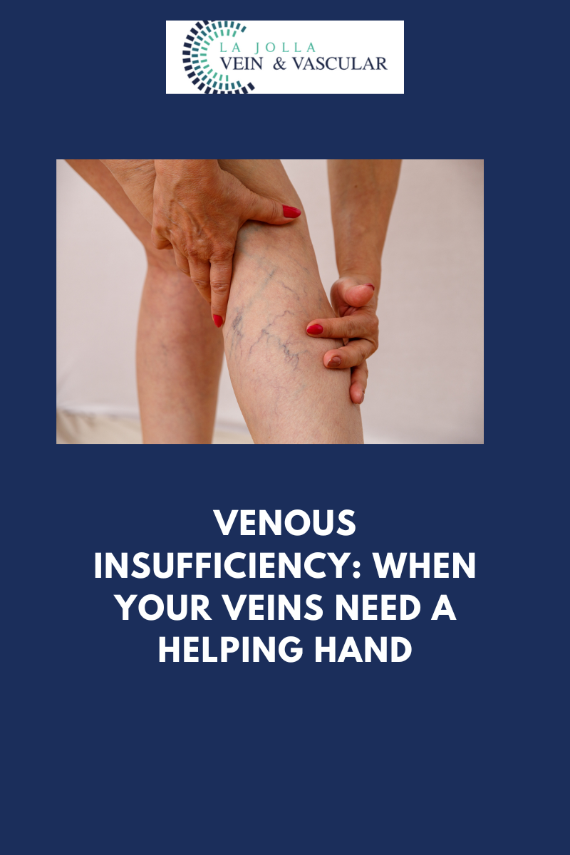
Venous reflux disease, sometimes referred to as venous stasis, venous insufficiency, or venous incompetence, stands as a multifaceted condition that intricately affects the veins in the legs. In this article, we delve into the intricacies of venous reflux disease, elucidating its causes and symptoms, and shedding light on the progressive nature of this ailment. We’ll also delve into the indispensable role played by ultrasound technology in the diagnostic process and the formulation of tailor-made treatment plans.

Unraveling Venous Reflux

At the core of venous reflux disease lies the concept of ‘leaky valves’ within the leg veins. These valves, entrusted with the responsibility of maintaining the smooth flow of blood, may falter, permitting blood to flow backward (a condition known as reflux) rather than progressing toward the heart. Venous reflux has the potential to manifest in both deep and superficial leg veins, subsequently influencing the efficiency of blood circulation.

Demystifying the Anatomy of Reflux

Within the realm of leg veins, there exist two primary categories: deep and superficial. The deep veins, nestling within the muscular tissue, shoulder the responsibility of transporting the lion’s share of blood from the legs back to the heart. Conversely, superficial veins are situated just beneath the skin, external to the muscle. Among the pivotal players in the realm of superficial veins are the Great Saphenous Vein (GSV), meandering through the thigh and calf, and the small saphenous vein, traversing the back of the calf.

The Ramifications of Leaky Valves

Under normal circumstances, one-way valves present in leg veins facilitate the upward flow of blood against the force of gravity, aided by the rhythmic contractions of calf muscles. However, when these valves succumb to leakage, the flow of blood reverses, leading to the accumulation of blood in the lower legs. This condition is accompanied by a medley of distressing symptoms, including the sensation of heavy legs, pain, fatigue, ankle swelling, and even restlessness in the legs during the night. As time unfolds, venous reflux disease can advance, resulting in skin changes such as darkening, dryness, itching, and the potential emergence of venous leg ulcers.

The Role of Ultrasound in Diagnosis

The diagnosis of venous reflux disease necessitates specialized tools, with ultrasound technology taking center stage. Not all vein-related issues are discernible to the naked eye, as many originate from veins concealed beneath the skin’s surface. Ultrasound examinations grant healthcare professionals valuable insights into the direction of blood flow, the functionality of valves, and the presence of any obstructions or scars within the veins.

A Tailored Approach to Treatment

Effectively addressing venous reflux disease requires a strategic and customized approach, recognizing the unique characteristics of each patient’s condition. The treatment process generally encompasses three fundamental steps:

Step 1: Rectifying Underlying Reflux

The initial focal point revolves around tackling the root cause—venous reflux. This is achieved by targeting the saphenous veins, which often serve as the epicenter of the issue. Cutting-edge vein ablation procedures, including radiofrequency ablation, laser ablation, mechanico-chemical ablation (MOCA), and Varithena Foam, are deployed to restore the standard flow of blood.

Step 2: Combating Varicose Veins

Once the underlying reflux has been successfully addressed, the focus shifts to addressing varicose veins. Techniques like foam sclerotherapy, involving the injection of a foamed medication, or minimally invasive removal methods, are utilized to eliminate bulging veins.

Step 3: Managing Spider Veins

For those desiring cosmetic enhancements, the treatment of spider veins through sclerotherapy is available. Although primarily cosmetic, this step serves to conclude the all-encompassing treatment journey.

Venous reflux disease presents as a multifaceted condition that demands specialized care for effective management. Our comprehensive approach encompasses state-of-the-art diagnostics, cutting-edge treatments, and patient-tailored care to address the various facets of this ailment. Through our expertise and unwavering dedication, our aim is to deliver transformative outcomes, ultimately enhancing the health and quality of life of our patients. If you are prepared to embark on the path to healthier veins, do not hesitate to reach out to us to take the first stride toward comprehensive vein and vascular wellness.

“Bringing Experts Together for Unparalleled Vein and Vascular Care”

La Jolla Vein & Vascular (formerly La Jolla Vein Care) is committed to bringing experts together for unparalleled vein and vascular care. 

Nisha Bunke, MD, Sarah Lucas, MD, and Amanda Steinberger, MD are specialists who combine their experience and expertise to offer world-class vascular care. 

Our accredited center is also a nationally known teaching site and center of excellence. 

For more information on treatments and to book a consultation, please give our office a call at 858-550-0330. 

 For a deeper dive into vein and vascular care, please check out our Youtube Channel at this link, and our website https://ljvascular.com

For more information on varicose veins and eliminating underlying venous insufficiency,

Please follow our social media Instagram Profile and Tik Tok Profile for more fun videos and educational information. 

For more blogs and educational content, please check out our clinic’s blog posts

Venous Insufficiency: When Your Veins Need a Helping Hand2023-10-11T16:30:21-07:00

Venous Duplex Ultrasound: Peering Beneath the Surface for Vascular Clarity

2023-10-11T16:20:49-07:00

Venous Duplex Ultrasound: Peering Beneath the Surface for Vascular Clarity

In the realm of understanding and diagnosing vein and vascular diseases, cutting-edge medical technology plays a pivotal role in uncovering the mysteries hidden beneath the surface. Among the most invaluable tools in this domain is Duplex Ultrasound. In this blog post, we embark on a journey to demystify Duplex Ultrasound, shedding light on its applications, advantages, and the pivotal role it plays in diagnosing and managing vein and vascular diseases.

Decoding the Enigma of Duplex Ultrasound

Duplex ultrasound is a specialized imaging technique that ingeniously combines two indispensable components: the traditional ultrasound (referred to as B-mode) and Doppler ultrasound. This amalgamation of technologies empowers healthcare professionals to not only visualize the structure of blood vessels but also assess the flow and speed of blood coursing within them.

The Versatility of Duplex Ultrasound

Duplex Ultrasound emerges as a versatile diagnostic tool, serving a multitude of purposes. Let’s navigate through some of its primary applications and advantages:

  • Detecting Vein Diseases: Duplex Ultrasound assumes a pivotal role in pinpointing various vein conditions, including varicose veins, deep vein thrombosis (DVT), and chronic venous insufficiency. By providing a visual portrayal of veins and scrutinizing blood flow, healthcare practitioners can identify irregularities and formulate precise diagnoses.
  • Assessing Arterial Conditions: Arterial maladies like peripheral arterial disease (PAD) and carotid artery disease yield to effective evaluation through the utilization of Duplex Ultrasound. This tool aids in recognizing arterial constriction, plaque accumulation, and blood flow obstructions, thereby contributing to timely medical interventions.
  • Guiding Interventional Procedures: Duplex Ultrasound emerges as invaluable for steering minimally invasive vascular interventions such as angioplasty and stent placement. Real-time imaging empowers physicians to navigate catheters and instruments with precision, thus addressing vascular blockages effectively.
  • Monitoring Blood Flow: The continual monitoring of blood flow stands as a critical necessity in numerous clinical scenarios. Duplex Ultrasound caters to this requirement, facilitating healthcare providers in evaluating the efficacy of treatments and tracking the progression of diseases.
  • Non-Invasive and Painless: One of the notable merits of Duplex Ultrasound lies in its non-invasive and painless nature. Patients encounter no need for special preparations, and there are no needles or exposure to radiation involved. This renders it a secure and well-tolerated imaging modality.
  • Immediate Results: Duplex Ultrasound yields real-time results, furnishing the advantage of swift assessments and instant feedback for both healthcare practitioners and patients alike. This expedites the diagnostic process and streamlines the decision-making procedure.
  • Risk Assessment: The application of Duplex Ultrasound extends to evaluating the risk of potential complications, such as blood clots or aneurysms. Early detection paves the way for preventive measures to mitigate these risks effectively.

The Procedure

During a Duplex Ultrasound procedure, a proficient technician, often referred to as a sonographer, administers a special gel to the skin covering the targeted area for examination. This gel serves a dual purpose by facilitating the transmission of ultrasound waves while preventing any interference from air. Subsequently, a handheld device called a transducer is gently maneuvered over the skin’s surface.

The transducer emits high-frequency sound waves that bounce off the blood vessels and return as echoes. These echoes are then transformed into images and Doppler signals in color-coded fashion, presenting a holistic view of the blood vessels’ architecture and blood flow patterns.

Duplex Ultrasound emerges as a remarkable diagnostic tool that empowers healthcare providers to delve beneath the surface, unraveling crucial insights about vein and vascular diseases. Its adaptability, non-invasive attributes, and real-time imaging capabilities render it an indispensable asset in the diagnosis and management of a wide spectrum of vascular conditions.

If you or someone you know is grappling with symptoms related to vein or vascular disease, seeking guidance from a healthcare provider who harnesses the power of Duplex Ultrasound can prove to be invaluable. Such consultations can provide vital insights and steer you towards the most suitable treatment and care plan.

“Bringing Experts Together for Unparalleled Vein and Vascular Care”

La Jolla Vein & Vascular (formerly La Jolla Vein Care) is committed to bringing experts together for unparalleled vein and vascular care. 

Nisha Bunke, MD, Sarah Lucas, MD, and Amanda Steinberger, MD are specialists who combine their experience and expertise to offer world-class vascular care. 

Our accredited center is also a nationally known teaching site and center of excellence. 

For more information on treatments and to book a consultation, please give our office a call at 858-550-0330. 

 For a deeper dive into vein and vascular care, please check out our Youtube Channel at this link, and our website https://ljvascular.com

For more information on varicose veins and eliminating underlying venous insufficiency,

Please follow our social media Instagram Profile and Tik Tok Profile for more fun videos and educational information. 

For more blogs a


Venous Duplex Ultrasound: Peering Beneath the Surface for Vascular Clarity2023-10-11T16:20:49-07:00

Unlocking Vital Insights: Understanding the Complex World of Vascular Disease

2023-10-11T16:13:10-07:00

Unlocking Vital Insights: Understanding the Complex World of Vascular Disease

Vascular disease is a term that may initially appear intricate and intimidating, but its ramifications on your health and overall well-being are anything but trivial. In this blog post, our aim is to demystify vascular disease by deconstructing what it encompasses, how it exerts its influence on your body, and why comprehending it is paramount for your holistic health. So, let’s peel back the layers and shed light on this vital facet of your vascular system.

Deciphering Vascular Disease

At its essence, vascular disease pertains to an array of conditions that impact the intricate network of blood vessels coursing through your body. These blood vessels can be broadly classified into two primary categories: arteries and veins. Arteries are responsible for ferrying oxygen-rich blood from your heart to all corners of your body, while veins work diligently to return blood back to your heart and lungs for replenishing with life-giving oxygen.

Common Manifestations of Vascular Disease

To grasp the concept of vascular disease, it’s imperative to acquaint oneself with the various forms it can assume and the ways in which it can influence your well-being. Here are some prevalent types of vascular diseases:

Arterial Disease: This category encompasses conditions like Peripheral Arterial Disease (PAD) and Carotid Artery Disease. For instance, PAD affects the arteries in your legs, often leading to reduced blood flow and discomfort. On the other hand, Carotid artery disease is linked to the arteries in your neck and can elevate the risk of stroke.

Venous Disease: Venous diseases encompass issues such as Varicose Veins, Deep Vein Thrombosis (DVT), and Chronic Venous Insufficiency. Varicose veins are visibly swollen and twisted veins, while DVT is a blood clot in a deep vein, typically occurring in the legs. Chronic Venous Insufficiency results from weakened valves in the veins, causing blood to pool in the legs.

Abdominal Aortic Aneurysm (AAA): This condition involves the expansion or ballooning of the abdominal aorta, which can lead to life-threatening complications should it rupture.

The Significance of Understanding Vascular Disease

Now that we’ve demystified the essence of vascular disease, let’s delve into why it is vital to comprehend this intricate subject for the sake of your health and overall well-being:

Early Detection and Prevention: Knowledge is a potent tool when it comes to vascular disease. Familiarity with its telltale signs and symptoms facilitates early detection, enabling proactive measures to avert complications. Early intervention can dramatically enhance outcomes.

Risk Factors: Vascular diseases frequently share common risk factors, such as smoking, high blood pressure, elevated cholesterol levels, obesity, and a sedentary lifestyle. By understanding these risk factors, you can take steps to mitigate them and diminish your chances of developing vascular diseases.

Lifestyle Modifications: Awareness of vascular disease can serve as a catalyst for making healthier lifestyle choices. This encompasses bidding farewell to smoking, embracing a consistent exercise regimen, and adhering to a heart-healthy diet, all of which contribute to the enhancement of your vascular health.

Treatment Options: Grasping the intricacies of vascular disease opens the gateway to a multitude of treatment choices. Depending on the type and severity of the condition, treatment options can span the spectrum from lifestyle adjustments and medications to minimally invasive procedures and surgical interventions. Being well-informed empowers you to make sound decisions regarding your health.

Enhanced Quality of Life: Vascular diseases can exert a substantial impact on your quality of life, subjecting you to discomfort, pain, and constraints in your daily activities. By comprehending these conditions, you can actively seek appropriate treatment and regain control over your well-being.

The process of unraveling the mysteries of vascular disease serves as the initial step toward taking command of your vascular health. Understanding what vascular disease entails, recognizing its common manifestations, and appreciating the significance of this knowledge empowers you to make informed choices regarding your health. Early detection, prevention, and lifestyle adaptations are pivotal keys to sustaining a healthy vascular system and a flourishing overall well-being.

If you harbor concerns or suspect that you may be at risk for vascular disease, do not hesitate to seek counsel from a healthcare provider. Your vascular health is an integral component of your overall well-being, and knowledge stands as the key to ensuring a brighter, healthier future.

“Bringing Experts Together for Unparalleled Vein and Vascular Care”

La Jolla Vein & Vascular (formerly La Jolla Vein Care) is committed to bringing experts together for unparalleled vein and vascular care. 

Nisha Bunke, MD, Sarah Lucas, MD, and Amanda Steinberger, MD are specialists who combine their experience and expertise to offer world-class vascular care. 

Our accredited center is also a nationally known teaching site and center of excellence. 

For more information on treatments and to book a consultation, please give our office a call at 858-550-0330. 

 For a deeper dive into vein and vascular care, please check out our Youtube Channel at this link, and our website https://ljvascular.com

For more information on varicose veins and eliminating underlying venous insufficiency,

Please follow our social media Instagram Profile and Tik Tok Profile for more fun videos and educational information. 

For more blogs and educational content, please check out our clinic’s blog posts

Unlocking Vital Insights: Understanding the Complex World of Vascular Disease2023-10-11T16:13:10-07:00

Exploring Arterial Exams: Comprehensive Vascular Care at La Jolla Vein and Vascular

2023-10-11T16:01:37-07:00

Exploring Arterial Exams: Comprehensive Vascular Care at La Jolla Vein and Vascular

Exploring La Jolla Vein & Vascular Arterial Assessments and Vascular Diagnostic Center

When it comes to preserving vascular health, the keys are early identification and precise diagnosis. At our facility, we present an all-inclusive solution for vascular imaging and consultations with expert physicians, all conveniently under one roof. With our cutting-edge non-invasive vascular diagnostic center, we leverage advanced imaging technology to detect and evaluate vascular conditions affecting blood circulation within arteries and veins. This blog post will take you through the advantages and expertise of our vascular diagnostic center, the array of conditions we diagnose, and the range of ultrasound testing we provide.

The Perks of Our Vascular Diagnostic Center

Our dedication to your vascular well-being transcends conventional healthcare. We offer an array of advantages to ensure your comfort and convenience:

Non-Invasive Testing: Our vascular diagnostic center specializes in non-invasive testing methods, guaranteeing a comfortable experience for our patients.

Same-Day Imaging Appointments: We acknowledge the importance of swift diagnosis. Thus, we provide same-day imaging appointments to promptly address your vascular concerns.

Comprehensive Care Under One Roof: Bid farewell to the hassle of juggling multiple locations for imaging, diagnosis, and treatment. At our facility, you can undergo vascular imaging and consult with a healthcare provider all in one place.

Private, Tranquil Rooms: Your comfort takes precedence in our book. Our private examination rooms are thoughtfully designed to offer a serene environment for your vascular tests.

Conditions We Identify

Our vascular diagnostic center is equipped to identify a wide spectrum of vascular conditions, including, but not limited to:

Venous Ailments:

  • Varicose Veins
  • Chronic Venous Insufficiency
  • Deep Vein Thrombosis (DVT)
  • Carotid Artery Disease and Stroke (Transient Ischemic Attack or Stroke)*

Arterial Disorders:

  • Peripheral Arterial Disease (PAD)**
  • Abdominal Aortic Aneurysm (AAA)
  • Upper Extremity Arterial Disease

Unpacking Duplex Ultrasound

Duplex ultrasound stands as a powerful diagnostic tool deployed in our vascular diagnostic center. It melds Doppler flow data with traditional imaging (B-mode) to deliver a comprehensive picture of your blood vessels. Essential elements of duplex ultrasound encompass:

Visualization of Structure: Duplex ultrasound empowers us to visualize the structure of your blood vessels, including their diameter and any obstructions.

Assessment of Blood Flow: It ascertains the velocity of blood flow within your vessels, examines the direction of blood flow, and identifies any impediments or blood clots.

Valve Function: This technology also evaluates the functionality of valves within your veins and arteries.

Deep Vein Evaluation: Duplex ultrasound has the capability to visualize deep veins nestled within the muscles, furnishing invaluable data for diagnosing conditions like varicose veins.

Varieties of Ultrasound Testing on Offer

Our vascular diagnostic center extends a diverse array of ultrasound testing options, including:

Direct Testing (Duplex Imaging):

  • Venous Assessments for Deep Vein Thrombosis and Venous Reflux
  • Arterial Examinations for the Abdominal Aorta, Abdominal Aortic Aneurysm (AAA) Screening, and Carotid Duplex
  • Lower Extremity Duplex

Indirect Testing (Non-Imaging):

  • Arterial Testing like Ankle Brachial Index (ABI) with waveforms and Segmental pressures with waveforms (P&Ws) for the upper or lower extremity
  • Venous Reflux or Venous Insufficiency Evaluation

Our Duplex Ultrasound examination empowers us to visualize blood vessels that remain concealed to the naked eye, even when they’re tucked deep within the muscles. This examination plays a pivotal role in evaluating the underlying cause of varicose veins by identifying veins with malfunctioning valves and mapping out their anatomical structure, ultimately charting a path for a precise assessment and an effective treatment strategy.

Preparing for Your Examination

Getting ready for our ultrasound study is a straightforward affair. Just keep these pointers in mind:

No specific preparation is needed for this study.

On the day of your examination, avoid wearing compression stockings.

Stay hydrated to ensure the best possible experience.

Our vascular diagnostic center is steadfast in delivering top-tier vascular imaging services in a setting that is both convenient and comfortable. Through non-invasive testing, same-day appointments, and a comprehensive approach to vascular health, our goal is to guarantee your well-being and peace of mind. Entrust us with your vascular concerns, and let us guide you on the path to enhanced health.

“Bringing Experts Together for Unparalleled Vein and Vascular Care”

La Jolla Vein & Vascular (formerly La Jolla Vein Care) is committed to bringing experts together for unparalleled vein and vascular care. 

Nisha Bunke, MD, Sarah Lucas, MD, and Amanda Steinberger, MD are specialists who combine their experience and expertise to offer world-class vascular care. 

Our accredited center is also a nationally known teaching site and center of excellence. 

For more information on treatments and to book a consultation, please give our office a call at 858-550-0330. 

 For a deeper dive into vein and vascular care, please check out our Youtube Channel at this link, and our website https://ljvascular.com

For more information on varicose veins and eliminating underlying venous insufficiency,

Please follow our social media Instagram Profile and Tik Tok Profile for more fun videos and educational information. 

For more blogs and educational content, please check out our clinic’s blog posts

Exploring Arterial Exams: Comprehensive Vascular Care at La Jolla Vein and Vascular2023-10-11T16:01:37-07:00

Cracking the Code of PAD: Understanding Peripheral Artery Disease

2023-10-11T15:55:33-07:00

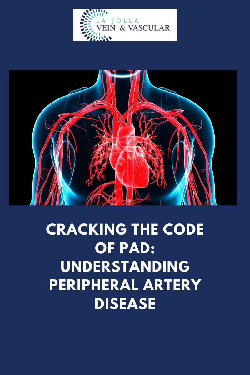
Cracking the Code of PAD: Understanding Peripheral Artery Disease

Peripheral Arterial Disease (PAD) can prove to be a formidable adversary, but with the right strategy for treatment, you can reclaim control over your life. The primary objectives of PAD treatment encompass the management of distressing symptoms, such as leg pain, and the thwarting of atherosclerosis progression, which in turn reduces the risk of grave health events like strokes and heart attacks. In this article, we will delve into a variety of treatment options for PAD, spanning from lifestyle adjustments to less invasive medical procedures.

Foundations of PAD Management: The Role of Lifestyle Modifications

Prior to delving into the specifics of various treatment choices, it is imperative to underscore the significance of lifestyle changes. Early intervention via lifestyle modifications can be remarkably effective in realizing the objectives of PAD treatment. Here are some pivotal steps:

  • Smoking Cessation: One of the most substantial measures you can take to arrest the advancement of PAD is to bid adieu to smoking. Smoking wreaks havoc on your blood vessels, exacerbating the condition.
  • Regular Physical Activity: Establishing a regimen of walking and supervised exercise training can remarkably ameliorate your PAD symptoms. Exercise fosters superior circulation and overall vascular well-being.

Less Invasive Procedures for Advanced PAD

Should your PAD reach a stage where lifestyle adjustments are no longer adequate, medical interventions may become imperative. Several less invasive procedures are at your disposal to address severe PAD:

Atherectomy

  • Atherectomy is a less invasive surgical procedure engineered to eliminate plaque from the arteries. Throughout the procedure, a catheter is introduced through a minor incision in the artery. The catheter possesses a chamber at its tip to collect the extracted plaque, ensuring thorough arterial cleansing. Typically performed under local anesthesia to minimize discomfort, the atherectomy may need to be repeated multiple times depending on the extent of plaque buildup to optimize blood flow.

Angioplasty (Percutaneous Transluminal Angioplasty – PTA)

  • Angioplasty is another minimally invasive procedure carried out using a catheter. This slender, flexible tube is directed by imaging to the narrowed segment of the artery. Upon reaching its destination, the catheter inflates a small balloon at its tip for a brief period. The inflation of the balloon applies pressure to the plaque, pushing it against the artery wall. This action widens the arteries, restoring normal blood flow.

Stenting

  • Stenting is frequently performed in conjunction with angioplasty and atherectomy. Over time, plaque buildup can inflict damage on artery walls, rendering them fragile and prone to collapsing even after plaque removal. Stenting entails the placement of a small mesh tube inside the artery to provide indispensable support. This tube acts as a scaffold, ensuring the artery remains unobstructed and permitting optimal blood flow.

The management of PAD involves a multi-pronged approach, commencing with lifestyle changes and escalating to minimally invasive procedures when necessary. Ceasing smoking and incorporating regular exercise serve as fundamental measures in the management of symptoms and the deceleration of atherosclerosis progression. If your condition has advanced, medical interventions like atherectomy, angioplasty, and stenting can provide relief and enhance your vascular health.

It is imperative to consult with your healthcare provider to discern the most appropriate treatment plan for your particular PAD condition. With the right strategy, you can efficaciously manage PAD, alleviate symptoms, and diminish the risk of grave cardiovascular incidents.

“Bringing Experts Together for Unparalleled Vein and Vascular Care”

La Jolla Vein & Vascular (formerly La Jolla Vein Care) is committed to bringing experts together for unparalleled vein and vascular care. 

Nisha Bunke, MD, Sarah Lucas, MD, and Amanda Steinberger, MD are specialists who combine their experience and expertise to offer world-class vascular care. 

Our accredited center is also a nationally known teaching site and center of excellence. 

For more information on treatments and to book a consultation, please give our office a call at 858-550-0330. 

 For a deeper dive into vein and vascular care, please check out our Youtube Channel at this link, and our website https://ljvascular.com

For more information on varicose veins and eliminating underlying venous insufficiency,

Please follow our social media Instagram Profile and Tik Tok Profile for more fun videos and educational information. 

For more blogs and educational content, please check out our clinic’s blog posts


Cracking the Code of PAD: Understanding Peripheral Artery Disease2023-10-11T15:55:33-07:00
Go to Top