22, 7, 2014

8 Warning Signs of Vein Disease: #5 Pregnancy Related Varicose or Spider Veins

2014-07-22T21:10:22-07:00
Pregnancy

Pregnant women should talk with their doctors about using medical compression therapy during pregnancy to reduce the risk of varicose veins and vein related issues.

8 Warning Signs of Vein Disease: #5 Varicose or Spider Veins, especially during or after pregnancy. During pregnancy, the amount of blood greatly increases throughout the body to almost double the normal volume, stretching leg veins far beyond their normal capacity.  Even though the visible signs of varicose veins may disappear after birth, the damage done to veins during pregnancy may be permanent and can cause pain and discomfort later in life. These problems may be avoided if compression therapy is prescribed during pregnancy. Women with a history of vein disease in their family or who experience swelling, pain or varicosities in the their legs during pregnancy are strongly urged to talk to their doctors about medical compression therapy or be evaluated by a vein specialist.

8 Warning Signs of Vein Disease: #5 Pregnancy Related Varicose or Spider Veins2014-07-22T21:10:22-07:00

Varicose Veins vs. Spider Veins

2021-11-05T13:08:51-07:00

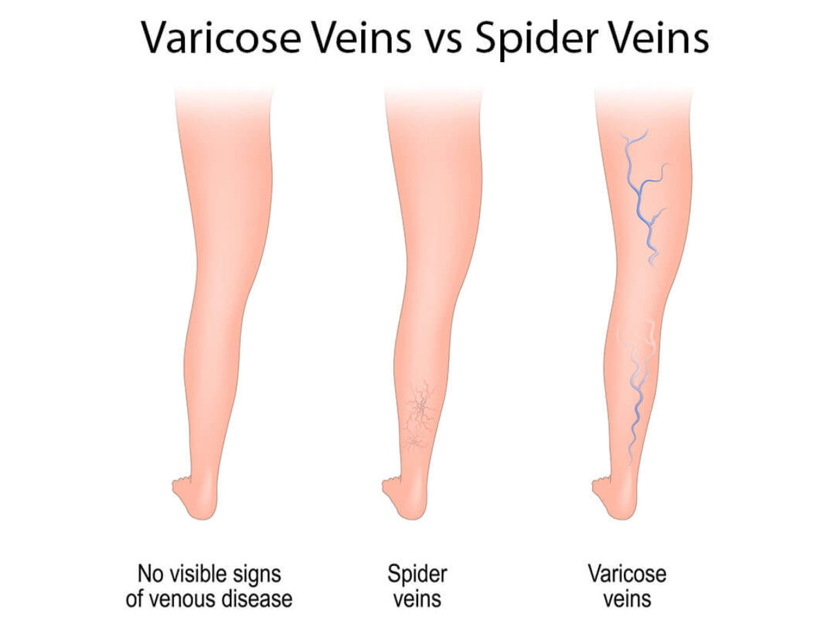
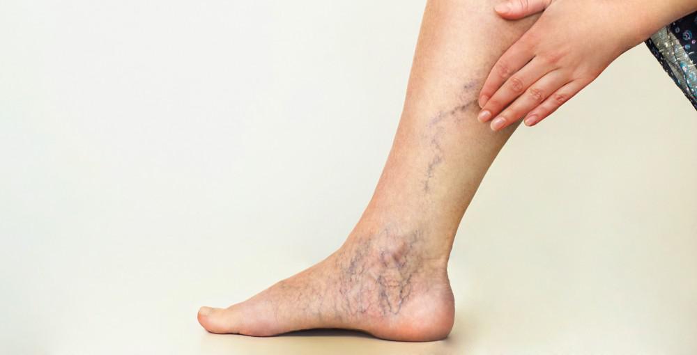
What is the difference between varicose veins and spider veins?  Are they the same thing? Spider veins and varicose veins both refer to dysfunctional, dilated leg veins but the main difference is the size of the veins. Spider veins are small, thread-like veins at the surface of the skin. They often appear in clusters or can have a ‘starburst’ or spider-like pattern. Varicose veins are larger veins that appear swollen, twisted cordlike veins that ‘bulge’ at the surface.

Both spider veins and varicose veins can cause pain and other symptoms like burning, aching, and throbbing. Both can be treated without surgery.

Slide 4_2

This image describes the difference between spider veins and varicose veins. Both are manifestations of unhealthy veins. Spider veins are essential, tiny varicose veins.

Varicose Veins vs. Spider Veins2021-11-05T13:08:51-07:00

How To Make Varicose Veins Vanish

2022-01-04T03:48:13-08:00

How To Make Varicose Veins Vanish Without Surgery

Physicians have many options for treating varicose veins. The choice often depends on the severity and cause of the problem. It’s important that patients are carefully evaluated so treatment can be tailored to individual needs.

In some cases, varicose veins may present a primarily cosmetic problem. In others, they may be a symptom of a more severe underlying circulatory condition that should be addressed by a vascular specialist.

Compression hosiery can be used to provide support and reduce varicose veins symptoms such as the aching and tiredness associated with but they cannot make varicose veins disappear.   There are no supplements or creams that can make varicose veins disappear. There are a variety of creams, lotions, and ointments that are advertised in newspapers and magazines to make veins disappear. There is no scientific evidence that these creams or lotions can make varicose and spider veins disappear. Once veins are dysfunctional, it is a mechanical problem and the valves cannot be repaired. But, they can be removed. There are many ways to accomplish this.

Sclerotherapy is used for smaller varicose veins and for spider veins, also known as telangiectasia. A fine needle is used to inject a chemical solution into the veins. The chemical causes the vein to collapse and eventually disappear. The procedure can be done in a doctor’s office and doesn’t require an anesthetic. Patients feel only a slight burning when the chemical is injected. Often several sessions are needed to treat the veins.

Foam sclerotherapy or ultrasound-guided foam sclerotherapy is used to treat large varicose veins.

Ambulatory Phlebectomy is an alternative to foam sclerotherapy, and involves making a series of tiny incisions along the length of the vein and gently removing the vein in sections using a hook. The procedure can be done on an outpatient basis and can be used for both large and small veins.

Radiofrequency ablation is a minimally invasive procedure involving using a catheter to introduce an electrode into the saphenous vein (great, small, or accessory saphenous veins). The energy from the electrode shrinks and seals off the vein. Nearby healthy veins take over the job of carrying blood from the legs.

Surgical ligation and stripping are rarely performed because the newer, minimally invasive procedures have replaced surgery for removing varicose veins.

For more information about treatment types and non-surgical methods to make varicose veins vanish, go to www.july01ljvascular.com. 

Before >> After

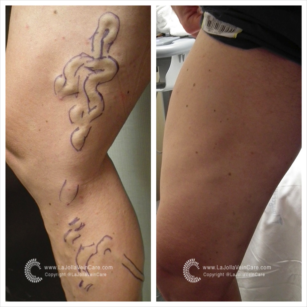
Before and After Pictures: Varicose Vein Treatment at La Jolla Vein Care. At La Jolla Vein Care, we utilize minimally invasive techniques to make varicose veins vanish- all without open surgery or general anesthesia.

How To Make Varicose Veins Vanish2022-01-04T03:48:13-08:00

How is Venous Reflux Diagnosed?

2021-11-05T11:48:32-07:00


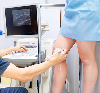
Venous duplex imaging uses ultrasound waves to create pictures.  La Jolla Vein Care utilizes state-of-the-art ultrasound scanners to image the veins beneath the surface of the skin, not visible to the naked eye. Duplex ultrasound imaging can identify if the vein is healthy, or if it is refluxing, or if there are any blood clots in the vein.

Duplex ultrasound combines Doppler flow information and conventional imaging information, sometimes called B-mode, to allow physicians to see the structure of your blood vessels. Duplex ultrasound shows how blood is flowing through your vessels and measures the speed of the flow of blood. It can also be useful to estimate the diameter of a blood vessel as well as the amount of obstruction, if any, in the blood vessel.  Conventional ultrasound uses painless sound waves higher than the human ear can detect that bounce off of blood vessels. A computer converts the sound waves into two-dimensional, black and white moving pictures called B-mode images.

Doppler ultrasound measures how sound waves reflect off of moving objects. A wand bounces short bursts of sound waves off of red blood cells and sends the information to a computer. When performing duplex ultrasound, your ultrasound technologist or physician uses the two forms of ultrasound together. The conventional ultrasound shows the structure of your blood vessels and the Doppler ultrasound shows the movement of your red blood cells through the vessels. Duplex ultrasound produces images that can be color coded to show physicians where your blood flow is severely blocked as well as the speed and direction of blood flow.  Venous reflux refers to back flow of blood across dysfunctional vein valves.  The direction of blood flow is detected by ultrasound.  This is measured in seconds.

US

La Jolla Vein Care image that shows reflux in the great saphenous vein. Duplex ultrasound combines Doppler flow information and conventional imaging information, sometimes called B-mode, to allow physicians to see the structure of your blood vessels.

How is Venous Reflux Diagnosed?2021-11-05T11:48:32-07:00

What is the Femoral Vein?

2022-01-03T13:29:29-08:00

The femoral vein is a large blood vessel of the leg that allows deoxygenated blood to travel to the heart and lungs to become oxygenated. It is located deep within the muscles of the thigh beginning just above the knee (at the adductor canal it is the continuation of the popliteal vein) and ends at the groin level (specifically, it ends at the inferior margin of the inguinal ligament where it becomes the external iliac vein.  It accompanies the femoral artery in the femoral sheath.  In this ultrasound image from La Jolla Vein Care, notice that the femoral vein runs along the same course as the femoral artery, which provides oxygenated blood from the heart and lungs to the rest of the body. The arteries and veins carry blood in opposite directions.  Ultrasound imaging detects the direction of blood flow and in this image, it femoral vein is ‘blue’ depicting blood flow moving toward the heart and the femoral artery is ‘red’ demonstrating blood flow away from the heart.

Ultra Sound detects Femoral Vein

Ultrasound image of the femoral vein and femoral artery. Ultrasound imaging detects the direction of blood flow and in this La Jolla Vein Care image, the femoral vein is ‘blue’ depicting blood flow moving toward the heart and the femoral artery is ‘red’ demonstrating blood flow away from the heart. Notice that the femoral vein and artery are located within the muscle. For orientation purposes, the skin is located at the top of the image.


What is the Femoral Vein?2022-01-03T13:29:29-08:00

Foam Treatment of Venous Malformations

2021-11-05T13:22:04-07:00
DSCF0086 copy

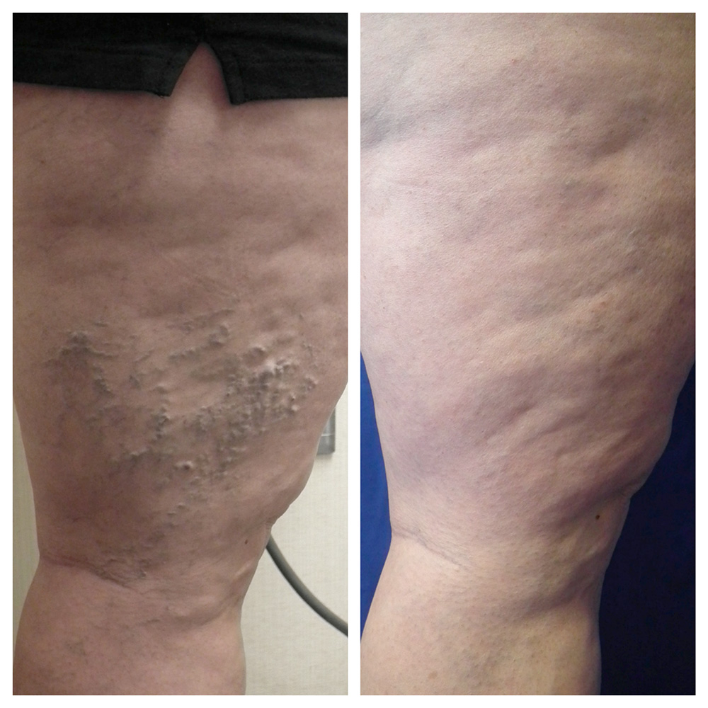
Venous malformation of the lateral leg before treatment. Venous malformations appear like abnormally dilated bluish vessels near the surface of the skin. They appear in clusters and look different than typical varicose veins. They often appear during childhood, as opposed to varicose veins.

Venous malformations comprise either superficial or deep veins that are abnormally formed and dilated.  Although they usually are present at birth, they may not be seen until years later into adolescence or even adulthood. The natural history of a venous malformation is slow, steady enlargement. However, events such as surgery, trauma, infection, or hormonal changes associated with puberty, pregnancy or menopause may cause rapid expansion.

At La Jolla Vein Care, we frequently evaluate and treat superficial venous malformations of the legs. Prior to treatment, it is important to have thorough diagnostic imaging, such as an ultrasound examination to ensure an accurate diagnosis of a pure venous malformation.  If an arterial connection (arterial venous malformation) is suspected on ultrasound evaluation, further imaging with CT or MR arteriogram may be necessary. Venous malformations can also occur in other syndromes, such as Klippel -Trenaunay syndrome and may involve the abnormal development of the deep veins.

Pure venous malformations can be treated without surgery. Foam sclerotherapy is a common treatment type.  Foam sclerotherapy uses a technique to inject a foamed sclerosing solution into the cluster of varicose veins, which will cause them to collapse and eventually dissolve.

DSCF1899

Venous malformation after treatment with ultrasound-guided foam sclerotherapy at La Jolla Vein Care. Treatment can reduce the symptoms associated with venous malformations such as leg pain, aching and throbbing.

Foam Treatment of Venous Malformations2021-11-05T13:22:04-07:00

8 Warning Signs of Vein Disease: #4 Swollen Ankles

2021-11-05T04:26:19-07:00
After

Ankle swelling related to varicose veins and venous insufficiency may be subtle, leaving indentations from socks.

Swollen Ankles at Night

Thick, swollen ankles are signs that blood or other fluid is congested in the leg and/or leg veins. Over time, damaged vein walls can become even more stretched out and permeable, allowing fluid and protein to filter from the veins into surrounding leg tissue. When you lie down at night, the pressure from gravity is equalized across your leg. Usually, vein related swelling (venous edema) becomes apparent later in the day or worse throughout the day and improved with leg elevation or overnight during sleep.  Often in the morning, there may be no swelling. But, as the day progresses gravity causes poo

Pooling of blood around the ankle in incompetent veins. Venous insufficiency is one of the most common causes of ankle swelling that worsens throughout the day. Sometimes, the swelling can be subtle, leaving indentations from sock lines. Or, it may cause the skin to feel firm, shiny, or puffy by the end of the day.

If you have swelling, you should discuss it with your healthcare provider. Venous insufficiency can be diagnosed with a venous duplex sonogram, which is a non-invasive study of the leg veins.

8 Warning Signs of Vein Disease: #4 Swollen Ankles2021-11-05T04:26:19-07:00

8 Warning Signs of Vein Disease: #3 Open Sores or Ulcers on the Lower Leg

2014-07-02T00:16:13-07:00
Before

Venous leg ulcers are the result of chronic venous insufficiency and venous hypertension, which causes the skin to break down.

When chronic venous insufficiency reaches its most serious point, ulcers may appear on the lower leg. These open ulcers are the result of blood leaking into the leg tissue and damaging the skin. Open sores need to be treated by a doctor immediately.

8 Warning Signs of Vein Disease: #3 Open Sores or Ulcers on the Lower Leg2014-07-02T00:16:13-07:00

30, 6, 2014

New Product: The Scat Belt Becoming a Favorite Exercise Companion

2022-01-04T04:33:14-08:00

Exercise is an important component of good vein health.  Exercise empties the veins of the legs and is beneficial to the cardiovascular system.  As a result, regular exercise, even a walking routine is beneficial to the leg veins and can help reduce leg symptoms. A new product was developed that keeps you safe during outdoor exercises, such as going on evening walks or hiking in areas that are less populated. The Scat Urban Belt, allows you to carry pepper spray, a super loud alarm, a smartphone, and even a bottle of water!  For more information, go to scatbelt.com.

Access pepper spray, an ear-piercing personal alarm, and smartphone within seconds – also carry a bottle of water.

scat-urban-indiegogo

The SCAT URBAN Belt is lightweight, comfortable, and provides peace of mind because you’re prepared and protected. The Urban comes in three great colors and two adjustable sizes for optimal fit.

New Product: The Scat Belt Becoming a Favorite Exercise Companion2022-01-04T04:33:14-08:00

8 Warning Signs of Vein Disease: #2 Discoloration of the Skin

2021-11-03T17:41:45-07:00

Discoloration of the Skin

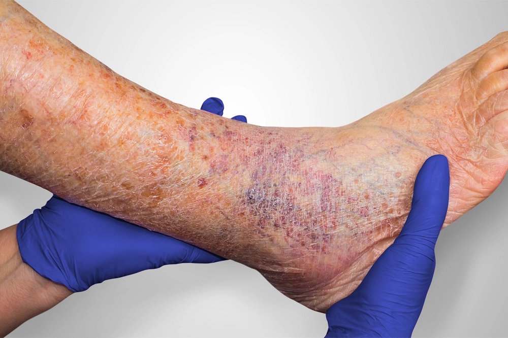
Over time, leakage of the blood into the area surrounding the veins can cause tissue to die. The resulting pooling of blood in the tissue causes a darkening of the skin. It is at this stage that the skin is actually stained by your own blood.  This is also known as venous stasis skin changes, skin hyperpigmentation, or venous stasis dermatitis because the skin is often inflamed, itchy, and eczema-like.  This is most frequently caused by venous reflux, an underlying condition that can be treated. When skin changes like these are present, the condition is termed chronic venous insufficiency. Over time, the skin condition may worsen and the skin may become darker, firm to touch, scaly, and itchy, and the skin may break down causing a venous leg ulcer.

8 Warning Signs of Vein Disease: #2 Discoloration of the Skin2021-11-03T17:41:45-07:00
Go to Top