15, 1, 2024

How do I alleviate Leg Pain?

2024-03-06T14:21:54-08:00

Alleviating Leg Pain: Your Complete Guide to Understanding and Finding Relief

Leg pain can stem from various sources, necessitating the identification of the underlying cause for suitable treatment. In this article, we’ll delve into the diverse factors contributing to leg pain, encompassing vascular issues like poor circulation, varicose veins, and blood clots, along with non-vascular causes such as injuries, arthritis, and low back discomfort. Discover more about the distinct types of leg pain and how seeking professional evaluation can lead to effective relief.

Understanding the Causes of Leg Pain

Vascular Factors:

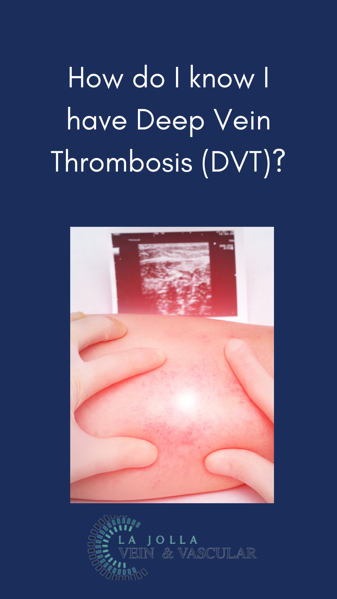
Blood Clots: Whether deep vein thrombosis (DVT) or superficial vein thrombosis, blood clots can induce leg pain. DVT might cause sudden swelling, pain, redness, and warmth in one leg. Superficial vein thrombosis typically affects varicose veins, causing pain, hardness, and a cord-like sensation beneath the skin.

Venous Reflux Disease: Characterized by leg pain, heaviness, and fatigue worsening with prolonged standing, this condition can be exacerbated by hormonal influences in women. Nighttime leg cramping and restlessness are common, often accompanied by visible signs like varicose veins, ankle swelling, and skin changes such as darkening or ulceration.

Non-Vascular Causes:

Injuries: Injuries to joints, bones, muscles, or ligaments can result in localized leg pain.

Arthritis: Arthritic conditions can lead to persistent leg discomfort.

Low Back Pain: Leg pain originating from the lower back can radiate down to the legs.

Understanding Peripheral Arterial Disease (PAD)

Peripheral arterial disease refers to insufficient arterial circulation in the legs, particularly during walking—and characterized by cramping, especially in the calf muscles. Typically, resting alleviates the discomfort, accompanied by additional signs such as pale toes and absent pulses in the feet.

Diagnosing and Managing Leg Pain

Diagnosing the origin involves a comprehensive approach:

Medical History: Healthcare providers gather information about your medical background, symptoms, and any risk factors.

Physical Examination: A thorough exam can unveil signs of vascular or non-vascular issues.

Non-Invasive Ultrasound Imaging: This imaging technique aids in visualizing blood flow and identifying potential vascular problems.

ABI Test: An Ankle-Brachial Index (ABI) test can screen for peripheral arterial disease.

Treatment Options and Relief

The encouraging news is that all these conditions are treatable, often through enhancing blood flow. Once diagnosed, suitable treatment plans, including lifestyle modifications, medications, minimally invasive procedures, and more, can be devised. Seeking professional guidance is crucial for tailoring the right treatment approach specific to your condition.

Seeking Relief 

If you’re grappling with leg pain, seeking professional evaluation is imperative. Understanding the root cause of your discomfort marks the initial step towards finding effective relief. Whether your leg pain originates from vascular or non-vascular sources, remember that viable treatment options exist. By consulting healthcare providers specializing in these conditions, you can restore comfort and mobility. Reach out to us at 858-550-0330 to schedule a diagnostic imaging session and consultation with our dedicated healthcare providers. Your journey towards pain-free legs commences with a simple call.

“Bringing Experts Together for Unparalleled Vein and Vascular Care”

La Jolla Vein & Vascular (formerly La Jolla Vein Care) is committed to bringing experts together for unparalleled vein and vascular care. 

Nisha Bunke, MD, Sarah Lucas, MD, and Amanda Steinberger, MD are specialists who combine their experience and expertise to offer world-class vascular care. 

Our accredited center is also a nationally known teaching site and center of excellence. 

For more information on treatments and to book a consultation, please give our office a call at 858-550-0330. 

 For a deeper dive into vein and vascular care, please check out our Youtube Channel at this link, and our website https://ljvascular.com

For more information on varicose veins and eliminating underlying venous insufficiency,

Please follow our social media Instagram Profile for more fun videos and educational information. 

For more blogs and educational content, please check out our clinic’s blog posts

How do I alleviate Leg Pain?2024-03-06T14:21:54-08:00

Understanding Venous Disease: Beyond Myths and Realities

2023-12-26T14:38:49-08:00

Understanding Venous Disease: Beyond Myths and Realities

Venous reflux disease, known by various names like venous stasis, venous insufficiency, or venous incompetence, is a complex condition that impacts the veins in the legs. This article aims to unravel the intricacies of venous reflux disease—its causes, symptoms, and its progressive nature. Additionally, we’ll delve into the pivotal role played by ultrasound technology in diagnosing this condition and formulating personalized treatment plans.

Deciphering Venous Reflux

Venous reflux disease is centered around the concept of ‘leaky valves’ within leg veins. These valves, responsible for maintaining proper blood flow, may malfunction, leading to the backward flow (reflux) of blood instead of its intended route towards the heart. This reflux can affect both deep and superficial leg veins, hampering efficient blood circulation.

Unveiling the Anatomy of Reflux

Leg veins can be categorized into two main types: deep and superficial. Deep veins, nestled within the muscles, primarily transport blood from the legs back to the heart. Meanwhile, superficial veins reside just beneath the skin, playing a significant role in this condition. Notably, the Great Saphenous Vein (GSV) coursing through the thigh and calf, along with the small saphenous vein along the back of the calf, are key players in superficial vein issues.

Impact of Leaky Valves

Normally, one-way valves in leg veins assist blood flow against gravity, aided by the contraction of calf muscles. When these valves malfunction, blood backflows, resulting in pooling in the lower legs. This leads to symptoms such as leg heaviness, pain, fatigue, ankle swelling, and even restless legs at night. Over time, this condition progresses, leading to skin changes like darkening, dryness, itching, and the potential development of venous leg ulcers.

Diagnosis through Ultrasound

The diagnosis of venous reflux disease demands specialized tools, with ultrasound technology being pivotal. Many vein issues aren’t visible to the naked eye, as they arise beneath the skin’s surface. Ultrasound examinations provide insights into blood flow direction, valve functionality, and the presence of blockages or scars in the veins.

Personalized Treatment Approach

Effectively managing venous reflux disease involves a tailored strategy for each patient. The treatment typically involves three key steps:

Step 1: Addressing Underlying Reflux

Treatment starts with addressing the root cause—venous reflux—often focusing on the saphenous veins. Innovative vein ablation procedures like radiofrequency ablation, laser ablation, mechanico-chemical ablation (MOCA), and Varithena Foam are employed to restore proper blood flow.

Step 2: Handling Varicose Veins

Once the underlying reflux is resolved, the focus shifts to varicose veins. Foam sclerotherapy or minimally invasive removal methods are employed to eliminate bulging veins.

Step 3: Managing Spider Veins

For those seeking cosmetic enhancement, spider vein treatment through sclerotherapy completes the comprehensive treatment journey.

Venous reflux disease demands specialized care for effective management. Our approach encompasses cutting-edge diagnostics, advanced treatments, and personalized care to address the diverse aspects of this condition. Through our expertise and commitment, we strive to provide transformative outcomes that enhance both the health and quality of life of our patients. If you’re ready to embark on the journey towards healthier veins, reach out to us to take the first step towards comprehensive vein and vascular wellness.

“Bringing Experts Together for Unparalleled Vein and Vascular Care”

La Jolla Vein & Vascular (formerly La Jolla Vein Care) is committed to bringing experts together for unparalleled vein and vascular care. 

Nisha Bunke, MD, Sarah Lucas, MD, and Amanda Steinberger, MD are specialists who combine their experience and expertise to offer world-class vascular care. 

Our accredited center is also a nationally known teaching site and center of excellence. 

For more information on treatments and to book a consultation, please give our office a call at 858-550-0330. 

 For a deeper dive into vein and vascular care, please check out our Youtube Channel at this link, and our website https://ljvascular.com

For more information on varicose veins and eliminating underlying venous insufficiency,

Please follow our social media Instagram Profile for more fun videos and educational information. 

For more blogs and educational content, please check out our clinic’s blog posts


Understanding Venous Disease: Beyond Myths and Realities2023-12-26T14:38:49-08:00

29, 12, 2023

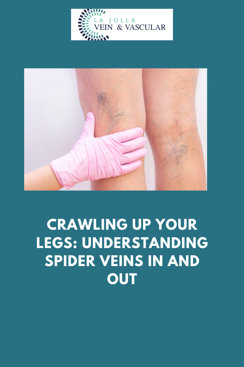
Spider veins: unmasking causes, solutions, and self confidence

2023-10-11T18:26:27-07:00

Spider veins: unmasking causes, solutions, and self confidence

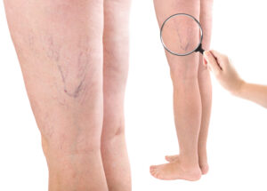
spider veins

Spider veins, those delicate veins with their intricate web-like patterns on the skin’s surface, are often viewed as primarily a cosmetic concern by many. However, beneath their aesthetic appearance, these tiny veins can hold valuable clues about your overall vein health. In this article, we embark on a journey into the world of spider veins, exploring their origins, symptoms, and the most effective treatment options available. Let’s unravel the truth about these small veins and discover how to manage them effectively.

Unraveling the Mystery of Spider Veins

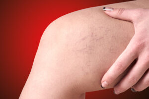
Scientifically known as telangiectasias, spider veins appear as fine, thread-like veins that create delicate patterns just below the skin’s surface. They come in various colors, including red, blue, and purple. While they might seem like a minor issue, spider veins can reveal more about your vein health than you might think.

The Connection to Feeder Veins and Venous Insufficiency

Although spider veins themselves are not typically considered a major health concern, they can be associated with less visible feeder veins or reticular veins. These feeder veins are often linked to larger veins and may provide clues about venous insufficiency, a condition where veins struggle to efficiently return blood to the heart. For example, if spider veins appear in specific areas like the inner ankles or inner thighs, it could indicate an underlying issue with the saphenous vein. Medical professionals may recommend ultrasound examinations to precisely locate and effectively address the source of these veins.

Recognizing Common Symptoms

Spider veins can manifest differently among individuals. While some might consider them solely a cosmetic issue without any physical discomfort, others may experience symptoms like a burning sensation, throbbing, localized pain, itchiness, and, in more severe cases, even bleeding. Paying attention to these symptoms is essential for determining the appropriate steps to manage and treat spider veins.

Unmasking the Underlying Causes

spider veins

On the legs, spider veins often share a common cause with varicose veins: malfunctioning vein valves. When these valves fail to prevent blood from pooling within the veins, it results in their enlargement and elongation. On the other hand, spider veins in other areas, such as the face and chest, can be caused by factors like sun damage, hormonal fluctuations, or liver disease. Hormonal changes during pregnancy, the use of birth control, or hormone replacement therapy can weaken vein walls, making them more prone to the emergence of spider veins.

Identifying Those at Risk

Several factors can increase an individual’s vulnerability to developing spider veins. These risk factors include genetics, pregnancy, female gender, advancing age, obesity, hormonal imbalances, and extended periods of sitting or standing. Recognizing these risk factors can be valuable for both prevention and early intervention.

Effective Treatment Options

For those seeking relief, various treatment options are available. The most common method for addressing spider veins on the legs is sclerotherapy. During this procedure, a special liquid sclerosant medication is injected into the affected veins, causing them to close, scar, and gradually fade away. Typically, multiple sessions are recommended for optimal results. While lasers can also be used to target fine spider veins, sclerotherapy remains the preferred choice due to its effectiveness.

While spider veins may appear as a minor concern, they can offer insights into your vein health. Whether you’re concerned about their appearance or you’re experiencing discomfort, it’s crucial to understand their potential underlying causes and seek appropriate treatment. By addressing spider veins with early intervention and effective treatments like sclerotherapy, you can not only enhance their appearance but also promote better overall vein health. Remember, caring for your veins is an investment in your well-being, one that pays off in both comfort and confidence.

“Bringing Experts Together for Unparalleled Vein and Vascular Care”

La Jolla Vein & Vascular (formerly La Jolla Vein Care) is committed to bringing experts together for unparalleled vein and vascular care. 

Nisha Bunke, MD, Sarah Lucas, MD, and Amanda Steinberger, MD are specialists who combine their experience and expertise to offer world-class vascular care. 

Our accredited center is also a nationally known teaching site and center of excellence. 

For more information on treatments and to book a consultation, please give our office a call at 858-550-0330. 

 For a deeper dive into vein and vascular care, please check out our Youtube Channel at this link, and our website https://ljvascular.com

For more information on varicose veins and eliminating underlying venous insufficiency,

Please follow our social media Instagram Profile and Tik Tok Profile for more fun videos and educational information. 

For more blogs and educational content, please check out our clinic’s blog posts

Spider veins: unmasking causes, solutions, and self confidence2023-10-11T18:26:27-07:00

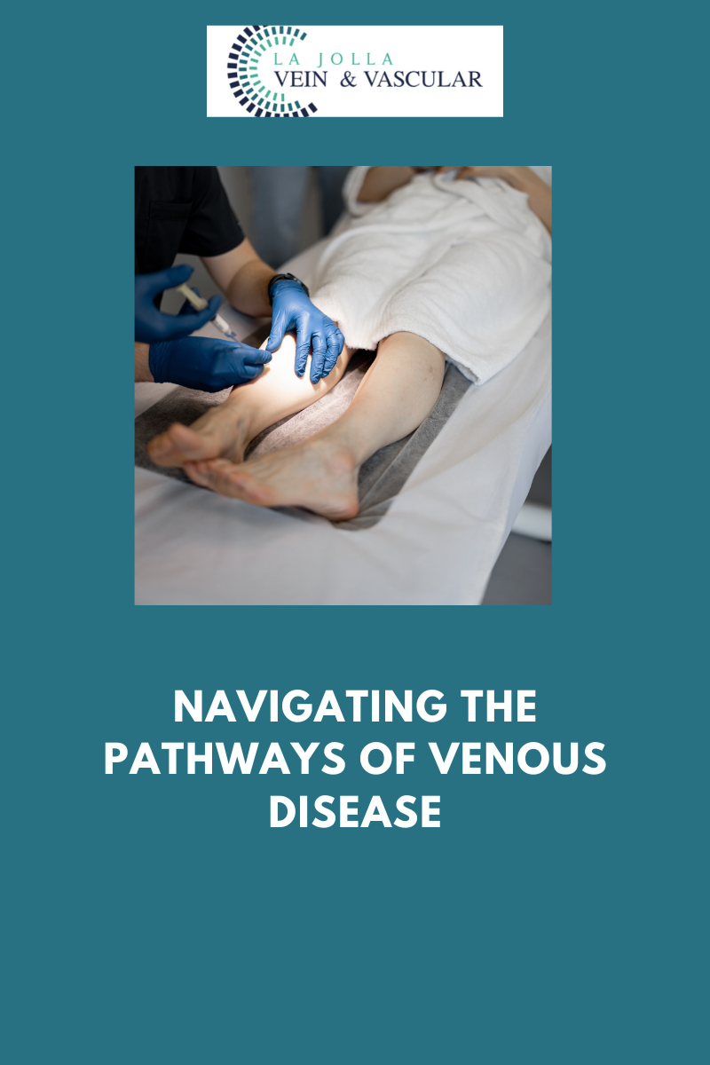
Navigating the Pathways of Venous Disease

2023-10-11T18:03:36-07:00

Navigating the Pathways of Venous Disease

Venous reflux disease, also known as venous stasis, venous insufficiency, or venous incompetence, is a multifaceted condition that affects the veins in the legs. In this article, we’ll take an in-depth look at venous reflux disease, including its underlying causes and the symptoms it presents. Additionally, we’ll delve into the progressive nature of this condition and the critical role played by ultrasound technology in both diagnosis and the formulation of personalized treatment plans.

Understanding Venous Reflux

At the core of venous reflux disease lies the concept of ‘leaky valves’ within the leg veins. These valves are responsible for maintaining the proper flow of blood, preventing it from flowing backward (reflux) towards the feet instead of moving upwards toward the heart. Venous reflux can occur in both the deep and superficial leg veins, significantly impacting blood circulation efficiency.

The Anatomy of Reflux

Leg veins can be categorized into two primary types: deep and superficial. Deep veins are located within the muscle and are responsible for carrying the majority of blood from the legs back to the heart. In contrast, superficial veins are situated just beneath the skin, outside the muscle. Within the realm of superficial veins, two key players are the Great Saphenous Vein (GSV), which courses through the thigh and calf, and the small saphenous vein, running along the back of the calf.

Implications of Leaky Valves

Normally, one-way valves in leg veins facilitate blood flow against gravity, with assistance from the contraction of calf muscles. When these valves become leaky, blood can flow backward, causing blood to pool in the lower legs. This condition manifests through various symptoms, such as leg heaviness, pain, fatigue, ankle swelling, and even restless legs at night. Over time, venous reflux disease can progress, leading to skin changes, including darkening, dryness, itching, and the potential development of venous leg ulcers.

Diagnosis Through Ultrasound

Accurate diagnosis of venous reflux disease necessitates specialized tools, with ultrasound technology taking the lead. Not all vein issues are visible to the naked eye, as many originate from veins beneath the skin’s surface. Ultrasound examinations offer a means to visualize the direction of blood flow, assess valve functionality, and detect the presence of blockages or scars within the veins.

Personalized Treatment Approaches

Effectively addressing venous reflux disease requires a tailored strategy for each patient. The treatment process typically comprises three key steps:

Step 1: Addressing Underlying Reflux

The primary focus is on addressing the root cause of the problem—venous reflux. This is typically achieved by targeting the saphenous veins, often the origin of the issue. Innovative vein ablation procedures, such as radiofrequency ablation, laser ablation, mechanico-chemical ablation (MOCA), and Varithena Foam, are utilized to restore proper blood flow.

Step 2: Managing Varicose Veins

After resolving the underlying reflux, attention shifts to treating varicose veins. This can involve foam sclerotherapy, a procedure using foamed medication injections, or minimally invasive removal methods to eliminate bulging veins.

Step 3: Treating Spider Veins

For those seeking cosmetic improvements, sclerotherapy is an option to address spider veins. While this step is primarily cosmetic, it contributes to the comprehensive treatment journey.

Venous reflux disease is a complex condition that requires specialized care for effective management. Our approach incorporates cutting-edge diagnostics, state-of-the-art treatments, and personalized patient care to comprehensively address the various facets of this condition. Through our expertise and unwavering commitment, we aim to provide transformative results that enhance both the health and quality of life for our patients. If you’re ready to embark on the path to healthier veins, don’t hesitate to reach out to us and take the initial step toward achieving comprehensive vein and vascular wellness.

“Bringing Experts Together for Unparalleled Vein and Vascular Care”

La Jolla Vein & Vascular (formerly La Jolla Vein Care) is committed to bringing experts together for unparalleled vein and vascular care. 

Nisha Bunke, MD, Sarah Lucas, MD, and Amanda Steinberger, MD are specialists who combine their experience and expertise to offer world-class vascular care. 

Our accredited center is also a nationally known teaching site and center of excellence. 

For more information on treatments and to book a consultation, please give our office a call at 858-550-0330. 

 For a deeper dive into vein and vascular care, please check out our Youtube Channel at this link, and our website https://ljvascular.com

For more information on varicose veins and eliminating underlying venous insufficiency,

Please follow our social media Instagram Profile and Tik Tok Profile for more fun videos and educational information. 

For more blogs and educational content, please check out our clinic’s blog posts

Navigating the Pathways of Venous Disease2023-10-11T18:03:36-07:00

Sclerotherapy for Spider Veins: Unveiling the Secrets to Flawless Legs

2023-10-11T17:53:10-07:00

Sclerotherapy for Spider Veins: Unveiling the Secrets to Flawless Legs

Have you grown weary of those persistent spider veins weaving their intricate patterns across your legs, causing you to hesitate before displaying your radiant skin? If that’s the case, rest assured that you’re not alone. The presence of spider veins and reticular veins is a common concern for many individuals. However, you need not fret, for there exists a magical remedy known as sclerotherapy that offers a means to bid farewell to these uninvited guests. In this blog post, we will reveal the captivating world of sclerotherapy, from the procedure itself to what you can anticipate on your treatment days.

Demystifying Sclerotherapy

Sclerotherapy stands as a well-established and remarkably effective treatment for spider veins and reticular veins, particularly when they grace your legs. The procedure involves the skillful injection of a specialized medication directly into the affected veins. This potent medication weaves its enchantment by compelling the veins to constrict and gradually fade into obscurity.

Why Choose Sclerotherapy?

You might be pondering why sclerotherapy is the favored choice of numerous vein specialists, surpassing alternative options such as laser treatments. The rationale behind this preference is as follows:

  • Minimal Discomfort: With newer solutions like AscleraTM, sclerotherapy has evolved to be more comfortable than ever. Patients can now bid farewell to the discomfort associated with older saline solutions.
  • Immediate Resumption of Activities: In contrast to certain treatments that demand downtime, sclerotherapy allows you to promptly resume your usual activities following the procedure.
  • Effective for Spider Veins: Sclerotherapy demonstrates particular efficacy when dealing with spider veins, which frequently have accompanying ‘feeder veins.’ It is these feeder veins that can be conveniently addressed through sclerotherapy, an aspect that remains unattended by laser treatments.

Anticipating the Sclerotherapy Treatment

If you find yourself contemplating sclerotherapy, it’s entirely natural to be curious about the treatment process. To ease your concerns, let’s embark on a step-by-step exploration of what you can envisage on your treatment days:

  • Consent and Preparation: Commencement is marked by the signing of a consent form. Subsequently, you’ll change into shorts thoughtfully provided by the medical facility to ensure unfettered access to the treatment area.
  • Skin Purification: The treatment area undergoes meticulous cleansing with alcohol, a precautionary measure to diminish the likelihood of infection.
  • The Injection: Employing a fine needle, the sclerosant medication is dexterously introduced into the veins. This medication is the secret elixir that shall diligently work its wonders on your spider veins.
  • Compression Stockings: Post-procedure, you will be assisted into compression stockings. These stockings emerge as vital players in augmenting the outcomes of sclerotherapy.
  • Walk it Off: To stimulate optimal circulation, you will be encouraged to embark on a 30-minute walk before settling into your vehicle. This seemingly simple step can wield a significant influence on the treatment’s triumph.

Sclerotherapy stands as the enchanted wand capable of making your spider veins vanish and in turn reinstating your self-assurance in the allure of your legs. With its minimal discomfort, immediate outcomes, and straightforward procedure, it is no surprise that sclerotherapy has secured its place as the favored choice of vein specialists. If you stand prepared to bid adieu to your spider veins, sclerotherapy may well be the enchanting solution that you’ve long been seeking.

“Bringing Experts Together for Unparalleled Vein and Vascular Care”

La Jolla Vein & Vascular (formerly La Jolla Vein Care) is committed to bringing experts together for unparalleled vein and vascular care. 

Nisha Bunke, MD, Sarah Lucas, MD, and Amanda Steinberger, MD are specialists who combine their experience and expertise to offer world-class vascular care. 

Our accredited center is also a nationally known teaching site and center of excellence. 

For more information on treatments and to book a consultation, please give our office a call at 858-550-0330. 

 For a deeper dive into vein and vascular care, please check out our Youtube Channel at this link, and our website https://ljvascular.com

For more information on varicose veins and eliminating underlying venous insufficiency,

Please follow our social media Instagram Profile and Tik Tok Profile for more fun videos and educational information. 

For more blogs and educational content, please check out our clinic’s blog posts

Sclerotherapy for Spider Veins: Unveiling the Secrets to Flawless Legs2023-10-11T17:53:10-07:00

Guardians of Life: Nurturing Arterial Health for a Vibrant Tomorrow

2023-10-11T17:12:31-07:00

Guardians of Life: Nurturing Arterial Health for a Vibrant Tomorrow

Within our intricate vascular system, arteries act as the vital highways, ensuring the efficient transportation of oxygen-rich blood throughout our bodies. This network plays a pivotal role in sustaining the proper functioning of our organs and tissues. However, the passage of time may present challenges to these indispensable thoroughfares, manifesting as issues like plaque buildup and blockages. In this blog post, we shall embark on a journey to understand the importance of arterial testing and screening, the diverse diagnostic methods available, and those individuals who should consider these crucial examinations.

Exploring Arterial Testing and Screening

Arterial testing and screening stand as foundational elements in the realm of vascular health assessment. These examinations furnish us with invaluable insights into the state of our arteries and serve as early detectors for concerns such as plaque accumulation, blockages, and reduced blood flow. Let us delve into some of the key diagnostic methods at our disposal:

  • Ankle-Brachial Index (ABI): ABI is a non-invasive evaluation that scrutinizes blood flow in both the legs and arms, employing inflatable pressure cuffs. This method yields vital information about the presence of arterial blockages and contributes to an overall assessment of arterial health.
  • Duplex Ultrasound: Duplex ultrasound emerges as a versatile instrument for scrutinizing the flow of blood through arteries and veins. This imaging tool encompasses various ultrasound tests, tailored to specific areas of interest:
    • Lower Extremity Arterial Study (LEA): This ultrasound zooms in on the arteries located in the lower legs, aiming to detect plaque buildup and potential blockages, often linked to atherosclerosis.
    • Upper Extremity Arterial Study (UEA): UEA takes a closer look at the arteries residing in the upper arms and neck, delivering insights into the state of arterial health in these regions.
    • Carotid Artery Ultrasound: This ultrasound focuses on assessing the carotid arteries in the neck, aiding in the identification or exclusion of blockages caused by plaque accumulation, which can elevate the risk of a stroke.
  • Abdominal Ultrasound: Abdominal ultrasound proves to be exceptionally valuable in the detection and evaluation of aneurysmal disease within the aorta and its branches. This includes the potentially perilous abdominal aortic aneurysm (AAA).

The Who and When of Arterial Screenings

Arterial screenings are essential for specific groups and individuals harboring risk factors. Consider the following guidelines for those who should contemplate these screenings:

  • Abdominal Aortic Aneurysm (AAA) Screening: According to recommendations by the U.S. Preventive Services Task Force (USPSTF) and the American Heart Association (AHA), men between the ages of 65 and 75 who have a history of smoking should contemplate undergoing ultrasound screening for AAA.
  • Peripheral Arterial Disease (PAD) Awareness: Given the elevated risk of heart attack and stroke associated with PAD, individuals falling within high-risk categories or those aged 55 and above should engage in conversations with their healthcare providers regarding the possibility of PAD screening. The importance of early diagnosis and treatment cannot be overstated.

The maintenance of arterial health forms the cornerstone of overall well-being, and the early detection of issues through testing and screening is a pivotal safeguard. Whether you find yourself concerned about your vascular health or you align with the recommended age and risk profiles, the deliberation of arterial screenings with your healthcare provider should be a priority. These essential examinations can provide you with the means to protect your arteries and reduce the risk of encountering severe cardiovascular complications.

“Bringing Experts Together for Unparalleled Vein and Vascular Care”

La Jolla Vein & Vascular (formerly La Jolla Vein Care) is committed to bringing experts together for unparalleled vein and vascular care. 

Nisha Bunke, MD, Sarah Lucas, MD, and Amanda Steinberger, MD are specialists who combine their experience and expertise to offer world-class vascular care. 

Our accredited center is also a nationally known teaching site and center of excellence. 

For more information on treatments and to book a consultation, please give our office a call at 858-550-0330. 

 For a deeper dive into vein and vascular care, please check out our Youtube Channel at this link, and our website https://ljvascular.com

For more information on varicose veins and eliminating underlying venous insufficiency,

Please follow our social media Instagram Profile and Tik Tok Profile for more fun videos and educational information. 

For more blogs and educational content, please check out our clinic’s blog posts

Guardians of Life: Nurturing Arterial Health for a Vibrant Tomorrow2023-10-11T17:12:31-07:00

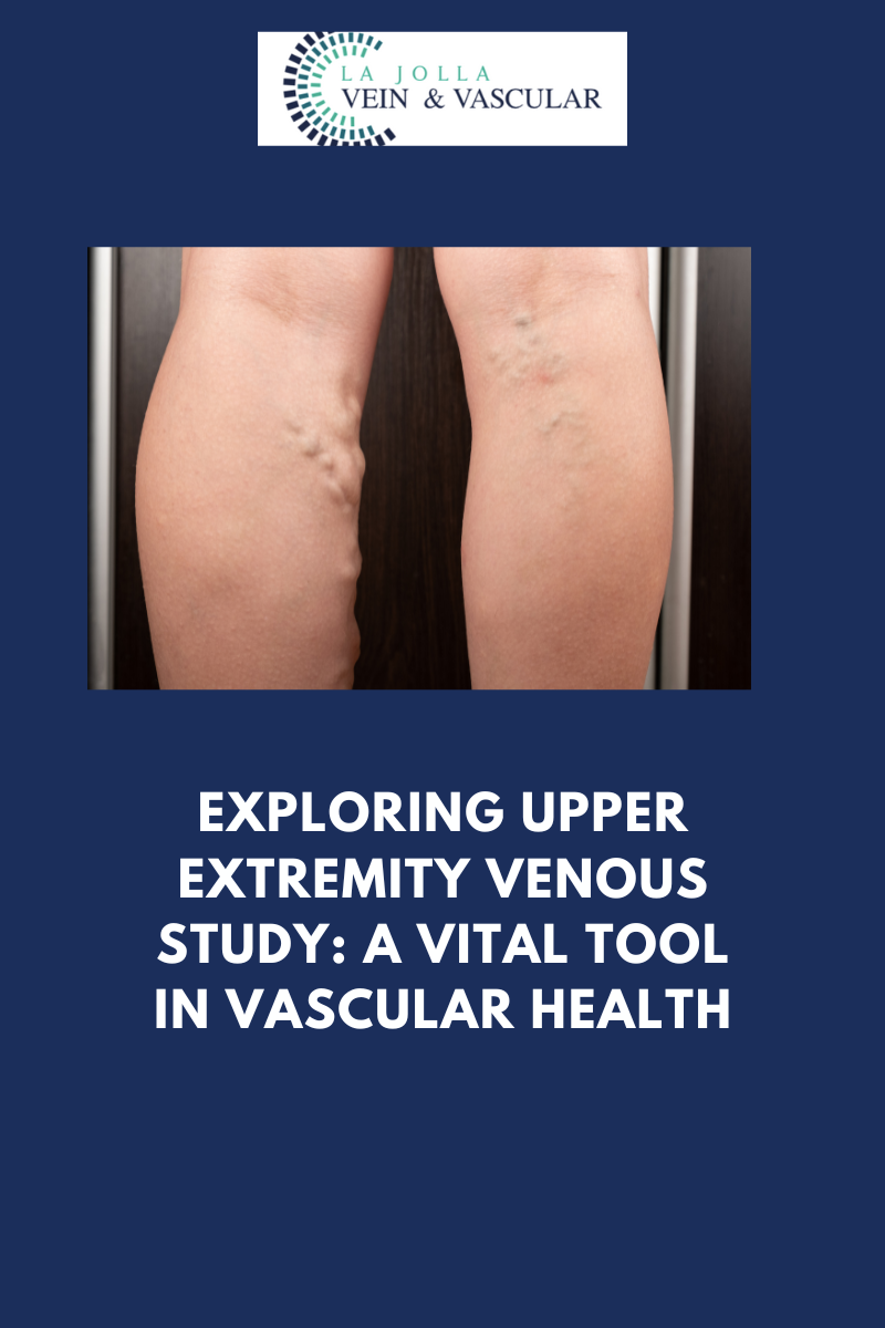
Exploring Upper Extremity Venous Study: A Vital Tool in Vascular Health

2023-10-11T17:03:31-07:00

Exploring Upper Extremity Venous Study: A Vital Tool in Vascular Health

While deep vein thrombosis (DVT) in the legs often garners significant attention, it’s of paramount importance not to disregard the upper extremities when evaluating vein health. Enter the Upper Extremity Venous Study, a specialized ultrasound examination that assumes a pivotal role in unveiling potential issues in the arms and neck. In this blog post, we will delve into the significance of this study, its objectives, and the role it plays in diagnosing arm-related concerns.

Deciphering the Upper Extremity Venous Study

The Upper Extremity Venous Study stands as a non-invasive imaging procedure with a dedicated focus on the veins residing in the upper arms and neck. Its primary mission is to thoroughly assess both the deep and superficial venous systems in these anatomical regions. This study takes center stage when an individual presents specific signs and symptoms, particularly pain and swelling in the arms, demanding careful investigation.

Why is an Upper Extremity Venous Study Conducted?

Numerous factors prompt healthcare providers to recommend the performance of an Upper Extremity Venous Study:

  • Arm Pain and Swelling: When a person experiences pain and swelling in one or both arms, especially if these symptoms remain unexplained or persistent, the Upper Extremity Venous Study proves invaluable in discerning the underlying causes.
  • Evaluation of Venous Health: Just as the lower extremities demand scrutiny for the presence of DVT, it is equally imperative to inspect the upper extremities for potential venous irregularities. This study is instrumental in identifying conditions such as deep vein thrombosis, superficial vein complications, and more.
  • Unexplained Symptoms: Occasionally, arm pain and swelling may manifest without an apparent cause. The Upper Extremity Venous Study, therefore, serves as a potent diagnostic tool to unveil concealed vascular concerns that might be at play.

The Procedure Unveiled

An Upper Extremity Venous Study is a procedure that adheres to a straightforward series of steps:

  • Preparation: For this examination, no specialized preparation is necessary. Patients can typically maintain their regular routines, including eating, drinking, and taking prescribed medications.
  • Application of Ultrasound Gel: A trained healthcare professional known as a sonographer will apply a specialized ultrasound gel to the skin in the areas of focus, which usually encompass the upper arms and neck.
  • Transducer Deployment: The procedure employs a handheld device known as a transducer, which emits high-frequency sound waves. These waves engage with the blood vessels within the arms and neck, generating echoes that are subsequently translated into images.
  • Real-Time Imaging: The transducer is maneuvered gently over the skin’s surface, affording real-time visualization of the veins in the upper extremities. This process unravels the veins’ structural details and facilitates an assessment of blood flow patterns.

The Upper Extremity Venous Study emerges as an invaluable instrument in the domain of vascular medicine. By comprehensively evaluating the deep and superficial venous systems within the arms and neck, it equips healthcare providers with the means to diagnose and address issues linked to arm pain, swelling, and other associated symptoms.

In the event that you, or someone within your purview, undergoes unexplained or persistent arm discomfort, seeking consultation with a healthcare provider and contemplating an Upper Extremity Venous Study can serve as an illuminating journey into the root causes. Early detection and intervention may pave the way for effective treatment and a brighter outlook for your arms.

“Bringing Experts Together for Unparalleled Vein and Vascular Care”

La Jolla Vein & Vascular (formerly La Jolla Vein Care) is committed to bringing experts together for unparalleled vein and vascular care. 

Nisha Bunke, MD, Sarah Lucas, MD, and Amanda Steinberger, MD are specialists who combine their experience and expertise to offer world-class vascular care. 

Our accredited center is also a nationally known teaching site and center of excellence. 

For more information on treatments and to book a consultation, please give our office a call at 858-550-0330. 

 For a deeper dive into vein and vascular care, please check out our Youtube Channel at this link, and our website https://ljvascular.com

For more information on varicose veins and eliminating underlying venous insufficiency,

Please follow our social media Instagram Profile and Tik Tok Profile for more fun videos and educational information. 

For more blogs and educational content, please check out our clinic’s blog posts

Exploring Upper Extremity Venous Study: A Vital Tool in Vascular Health2023-10-11T17:03:31-07:00

Deep Vein Thrombosis (DVT): Understanding the Silent Threat to Vascular Health

2023-10-11T16:50:51-07:00

Deep Vein Thrombosis (DVT): Understanding the Silent Threat to Vascular Health

Deep Vein Thrombosis, more commonly known as DVT, is a medical condition characterized by the formation of blood clots deep within the veins of the lower extremities. While these clots may remain concealed beneath the surface, the lurking danger they represent should never be underestimated. In this blog post, we embark on an in-depth exploration of DVT, unraveling its intricacies, recognizing the severity of this medical condition, elucidating the diagnostic process, and emphasizing the paramount importance of staying vigilant in its detection.

Deciphering Deep Vein Thrombosis (DVT)

Deep Vein Thrombosis, the ominous acronym of DVT, is a condition where blood clots form within the deep veins of the legs. The initial appearance of these clots might deceive, seemingly harmless; however, the true peril emerges when they venture into the bloodstream. If these clots journey to the lungs, they can give rise to a potentially life-threatening condition known as a pulmonary embolism.

The Pivotal Role of Duplex Ultrasound in Diagnosis

Diagnosing DVT stands as a pivotal stride towards averting its perilous consequences. This is where Duplex ultrasound, a priceless diagnostic instrument, assumes its critical role. Here is an insight into its functionality:

Experienced Vascular Sonographer: A proficient vascular sonographer serves as the linchpin in the diagnosis of DVT. Their expertise is the bedrock upon which accurate and reliable results are founded.

Duplex Ultrasound Examination: Duplex ultrasound amalgamates the prowess of traditional ultrasound, denoted as B-mode, with Doppler ultrasound. This fusion empowers healthcare providers to visualize the flow of blood and identify clots within the deep veins.

Indications for DVT Study: A DVT study is typically conducted when an individual presents with discernible signs and symptoms of lower extremity DVT. These symptoms often encompass leg pain and swelling, frequently accompanied by telltale signs like redness and warmth.

Routine Screening: In specific circumstances, such as post-vein procedures or other surgical interventions, routine DVT screening may be undertaken as a precautionary measure to identify clots before they manifest harm.

Preparation for the DVT Study

Fortunately, preparing for a DVT study is a straightforward endeavor, devoid of intricate steps. Here are the salient points to keep in mind:

No Special Preparation: In contrast to certain medical tests that mandate fasting or adherence to specific preparations, a DVT study typically necessitates no extraordinary measures.

Vigilance Beyond the Lower Limbs: The Upper Extremity Venous Study

Although DVT is conventionally associated with the lower extremities, it is imperative to recognize that blood clots can potentially take root elsewhere within the body. This includes the upper arms and neck. An Upper Extremity Venous Study is a diagnostic ultrasound procedure designed to investigate the upper arms and neck when a person exhibits symptoms such as pain and swelling in these regions.

Deep Vein Thrombosis epitomizes a grave medical condition, harboring the potential for consequences that may alter the course of a life. Timely diagnosis through Duplex ultrasound is the keystone of effective intervention and the prevention of complications, such as the ominous pulmonary embolism.

If you, or someone within your sphere of concern, experiences symptoms such as leg pain, swelling, or discomfort of obscure origin, it is imperative to promptly seek medical attention. Contemplating a DVT study can be the critical move that potentially saves a life. In the ever-present shadow of this concealed danger lurking within our veins, vigilance and early detection emerge as the unsung heroes.

“Bringing Experts Together for Unparalleled Vein and Vascular Care”

La Jolla Vein & Vascular (formerly La Jolla Vein Care) is committed to bringing experts together for unparalleled vein and vascular care. 

Nisha Bunke, MD, Sarah Lucas, MD, and Amanda Steinberger, MD are specialists who combine their experience and expertise to offer world-class vascular care. 

Our accredited center is also a nationally known teaching site and center of excellence. 

For more information on treatments and to book a consultation, please give our office a call at 858-550-0330. 

 For a deeper dive into vein and vascular care, please check out our Youtube Channel at this link, and our website https://ljvascular.com

For more information on varicose veins and eliminating underlying venous insufficiency,

Please follow our social media Instagram Profile and Tik Tok Profile for more fun videos and educational information. 

For more blogs and educational content, please check out our clinic’s blog posts

Deep Vein Thrombosis (DVT): Understanding the Silent Threat to Vascular Health2023-10-11T16:50:51-07:00

Vein Mapping: Navigating the Path to Improved Vascular Health

2023-10-11T16:41:15-07:00

Vein Mapping: Navigating the Path to Improved Vascular Health

vein mapping vein mapping

vein mapping

In the realm of diagnosing and addressing conditions related to veins, the importance of precise and comprehensive data cannot be overstated. Vein mapping ultrasound, often simply known as “vein mapping,” is an indispensable instrument within the domain of vascular medicine. In this blog post, we embark on an exploration of the pivotal role played by vein mapping ultrasound, its operational mechanisms, and the reasons it constitutes an indispensable preliminary step before embarking on any vein-related procedures.

The Significance of Vein Mapping

Vein mapping ultrasound fulfills a specific and vital role in the evaluation and management of issues linked to veins. Let’s delve deeper into its primary functions:

  • Unveiling Vein Anatomy: The moniker “vein mapping” is aptly chosen, as it serves as a detailed “roadmap” unveiling the intricate network of veins in the specified area. This visual map allows healthcare professionals to gain insight into the vein’s architecture, comprising critical factors such as size, location, and branching patterns.
  • Diameter Measurement: The diameters of veins are far from uniform and can exhibit variations at various points along a vein. Vein mapping ultrasound performs precise measurements at multiple sites. This information is pivotal for comprehending the scope of the condition and devising tailored treatment strategies.
  • Precise Evaluation: By synergizing the visual depiction of the veins with meticulous measurements, vein mapping ultrasound empowers healthcare providers to perform accurate evaluations of a wide spectrum of vein-related conditions. This includes conditions such as varicose veins, chronic venous insufficiency, and deep vein thrombosis.

The Imperativeness of Preparation

While the vein mapping ultrasound procedure itself is straightforward and devoid of discomfort, there are essential prerequisites to observe before undergoing the examination:

  • Steer Clear of Compression Stockings: It is imperative not to wear compression stockings on the same day as your examination. These garments can potentially hinder the ultrasound imaging process, adversely affecting the precision of the results.
  • Maintain Adequate Hydration: Adequate hydration is instrumental in securing optimal imaging results. Ensuring that you are adequately hydrated prior to the test contributes to obtaining crystal-clear images, thus facilitating healthcare providers in their accurate assessment of your veins.

The Procedure

During a vein mapping ultrasound, a proficient sonographer or vascular technician will oversee the procedure. Here’s an overview of what to expect:

Gel Application: The technician will gently apply a specialized ultrasound gel to the specific area under examination. This gel functions to transmit ultrasound waves effectively and ensures that there is no interference stemming from air between the transducer and your skin.

Transducer Utilization: The examination relies on the utilization of a handheld device known as a transducer. This transducer emits high-frequency sound waves that are directed towards the blood vessels. These waves bounce back as echoes, which are subsequently transformed into visual images disclosing the structure and measurements of the veins.

Vein Mapping: As the transducer is meticulously maneuvered across the surface of your skin, it generates a comprehensive visual “map” of the veins, detailing their dimensions and placements. This invaluable information is then recorded for further scrutiny and analysis.

Vein mapping ultrasound stands as an irreplaceable asset within the realm of vascular medicine. It empowers healthcare providers to craft an intricate blueprint of your veins, measure vein diameters, and perform precise evaluations of an array of vein-related conditions. This wealth of information serves as the cornerstone for crafting a personalized treatment plan tailored to your unique requirements.

If you are on the verge of a vein-related procedure or hold concerns regarding the health of your veins, comprehending the pivotal role of vein mapping ultrasound can serve as a beacon of assurance. It is an instrumental stride along the path towards healthier veins and an enhanced overall state of well-being.

“Bringing Experts Together for Unparalleled Vein and Vascular Care”

La Jolla Vein & Vascular (formerly La Jolla Vein Care) is committed to bringing experts together for unparalleled vein and vascular care. 

Nisha Bunke, MD, Sarah Lucas, MD, and Amanda Steinberger, MD are specialists who combine their experience and expertise to offer world-class vascular care. 

Our accredited center is also a nationally known teaching site and center of excellence. 

For more information on treatments and to book a consultation, please give our office a call at 858-550-0330. 

 For a deeper dive into vein and vascular care, please check out our Youtube Channel at this link, and our website https://ljvascular.com

For more information on varicose veins and eliminating underlying venous insufficiency,

Please follow our social media Instagram Profile and Tik Tok Profile for more fun videos and educational information. 

For more blogs and educational content, please check out our clinic’s blog posts

Vein Mapping: Navigating the Path to Improved Vascular Health2023-10-11T16:41:15-07:00

Venous Duplex Ultrasound: Peering Beneath the Surface for Vascular Clarity

2023-10-11T16:20:49-07:00

Venous Duplex Ultrasound: Peering Beneath the Surface for Vascular Clarity

In the realm of understanding and diagnosing vein and vascular diseases, cutting-edge medical technology plays a pivotal role in uncovering the mysteries hidden beneath the surface. Among the most invaluable tools in this domain is Duplex Ultrasound. In this blog post, we embark on a journey to demystify Duplex Ultrasound, shedding light on its applications, advantages, and the pivotal role it plays in diagnosing and managing vein and vascular diseases.

Decoding the Enigma of Duplex Ultrasound

Duplex ultrasound is a specialized imaging technique that ingeniously combines two indispensable components: the traditional ultrasound (referred to as B-mode) and Doppler ultrasound. This amalgamation of technologies empowers healthcare professionals to not only visualize the structure of blood vessels but also assess the flow and speed of blood coursing within them.

The Versatility of Duplex Ultrasound

Duplex Ultrasound emerges as a versatile diagnostic tool, serving a multitude of purposes. Let’s navigate through some of its primary applications and advantages:

  • Detecting Vein Diseases: Duplex Ultrasound assumes a pivotal role in pinpointing various vein conditions, including varicose veins, deep vein thrombosis (DVT), and chronic venous insufficiency. By providing a visual portrayal of veins and scrutinizing blood flow, healthcare practitioners can identify irregularities and formulate precise diagnoses.
  • Assessing Arterial Conditions: Arterial maladies like peripheral arterial disease (PAD) and carotid artery disease yield to effective evaluation through the utilization of Duplex Ultrasound. This tool aids in recognizing arterial constriction, plaque accumulation, and blood flow obstructions, thereby contributing to timely medical interventions.
  • Guiding Interventional Procedures: Duplex Ultrasound emerges as invaluable for steering minimally invasive vascular interventions such as angioplasty and stent placement. Real-time imaging empowers physicians to navigate catheters and instruments with precision, thus addressing vascular blockages effectively.
  • Monitoring Blood Flow: The continual monitoring of blood flow stands as a critical necessity in numerous clinical scenarios. Duplex Ultrasound caters to this requirement, facilitating healthcare providers in evaluating the efficacy of treatments and tracking the progression of diseases.
  • Non-Invasive and Painless: One of the notable merits of Duplex Ultrasound lies in its non-invasive and painless nature. Patients encounter no need for special preparations, and there are no needles or exposure to radiation involved. This renders it a secure and well-tolerated imaging modality.
  • Immediate Results: Duplex Ultrasound yields real-time results, furnishing the advantage of swift assessments and instant feedback for both healthcare practitioners and patients alike. This expedites the diagnostic process and streamlines the decision-making procedure.
  • Risk Assessment: The application of Duplex Ultrasound extends to evaluating the risk of potential complications, such as blood clots or aneurysms. Early detection paves the way for preventive measures to mitigate these risks effectively.

The Procedure

During a Duplex Ultrasound procedure, a proficient technician, often referred to as a sonographer, administers a special gel to the skin covering the targeted area for examination. This gel serves a dual purpose by facilitating the transmission of ultrasound waves while preventing any interference from air. Subsequently, a handheld device called a transducer is gently maneuvered over the skin’s surface.

The transducer emits high-frequency sound waves that bounce off the blood vessels and return as echoes. These echoes are then transformed into images and Doppler signals in color-coded fashion, presenting a holistic view of the blood vessels’ architecture and blood flow patterns.

Duplex Ultrasound emerges as a remarkable diagnostic tool that empowers healthcare providers to delve beneath the surface, unraveling crucial insights about vein and vascular diseases. Its adaptability, non-invasive attributes, and real-time imaging capabilities render it an indispensable asset in the diagnosis and management of a wide spectrum of vascular conditions.

If you or someone you know is grappling with symptoms related to vein or vascular disease, seeking guidance from a healthcare provider who harnesses the power of Duplex Ultrasound can prove to be invaluable. Such consultations can provide vital insights and steer you towards the most suitable treatment and care plan.

“Bringing Experts Together for Unparalleled Vein and Vascular Care”

La Jolla Vein & Vascular (formerly La Jolla Vein Care) is committed to bringing experts together for unparalleled vein and vascular care. 

Nisha Bunke, MD, Sarah Lucas, MD, and Amanda Steinberger, MD are specialists who combine their experience and expertise to offer world-class vascular care. 

Our accredited center is also a nationally known teaching site and center of excellence. 

For more information on treatments and to book a consultation, please give our office a call at 858-550-0330. 

 For a deeper dive into vein and vascular care, please check out our Youtube Channel at this link, and our website https://ljvascular.com

For more information on varicose veins and eliminating underlying venous insufficiency,

Please follow our social media Instagram Profile and Tik Tok Profile for more fun videos and educational information. 

For more blogs a


Venous Duplex Ultrasound: Peering Beneath the Surface for Vascular Clarity2023-10-11T16:20:49-07:00
Go to Top