5, 8, 2022

Hemorrhages & Varicose Veins

2022-07-19T17:31:08-07:00

Complications of Untreated Varicose Veins: Bleeding or Hemorrhage Caused by Vein Rupture


Untreated varicose veins are at a higher than usual risk of bleeding or spontaneous rupture, hemorrhage. Over time, varicose veins become larger, and the vein wall becomes weak and stretched out. These veins, which are already weak, are also under high pressure (because of venous reflux, or the ‘backflow’ and pooling of blood in these veins). As a result, the high pressure can cause the veins to spontaneously burst and bleed heavily. Because they are under high pressure, they bleed like an arterial bleed and patients describe the bleeding as ‘blood shooting across the room.’  


The varicose veins that are susceptible are veins closest to the surface of the skin.

Most patients describe that it occurs during or after a warm shower (warm water causes veins to relax and dilate, allowing more blood to pool within the veins) or during sleep. It is painless and patients report that they notice it because they feel something wet in bed.  Patients who are on blood thinners can lose large amounts of blood, especially if it occurs while they are sleeping. Some people have required blood transfusions. The small blue spider veins around the ankle are equally at risk of rupture as are the larger bulging veins.
hemorrhage

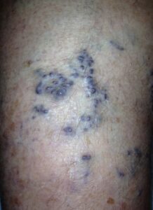
This is a patient who experienced spontaneous hemorrhage of their varicose veins. Notice the appearance of the blue, bulging veins that we describe as, ‘blue blebs.’ These veins are dilated, weak, and are close to the surface of the skin. The pooling blood within these veins causes high pressure, ultimately resulting in a spontaneous rupture or hemorrhage. The bleeding is rapid since varicose veins are under high pressure.  This is a common condition that we see at La Jolla Vein Care.


If someone you know has experienced bleeding from their varicose veins, they should be seen by a doctor. Treatment will prevent the veins from bursting again. This is a common condition that we see at La Jolla Vein Care.


For more information and to book a consultation, please give our office a call at 858-550-0330. 


For more information please check out our Youtube Channel at this link. 


Hemorrhages & Varicose Veins2022-07-19T17:31:08-07:00

Cellulitis & Varicose Veins

2022-07-19T17:21:50-07:00

Complications of Untreated Varicose Veins and Venous Insufficiency: Cellulitis

Cellulitis is a bacterial infection of the skin and tissues beneath the skin. Cellulitis infections can occur in the legs of people with untreated varicose veins and chronic venous insufficiency.  The reason for this is chronic venous insufficiency-which causes inflammation within the skin and underlying tissues. This inflammatory process causes the skin to become firm, eczema-like, dry, itchy, and fragile. Healthy skin acts as a barrier to bacteria, preventing infections. But, in chronic venous insufficiency, the skin is fragile and this barrier is susceptible to bacterial infections that can enter the tissues through cracks or breakages within the skin.  In chronic venous insufficiency, the skin around the ankles is most affected and infections can begin here.

Cellulitis usually begins as a small area of pain and redness on the skin. This area spreads to surrounding tissues, resulting in the typical signs of inflammation such as redness, swelling, warmth, and pain. Fever and chills may develop and the redness will spread affecting more of the leg. It is treated with antibiotics. If you believe you have signs of cellulitis, you need to contact your doctor right away as untreated cellulitis can spread rapidly.

This complication can be avoided by treating the varicose veins and eliminating underlying venous insufficiency, which will, in turn, reduce inflammation and improve the skin condition. 

For more information on varicose veins and eliminating underlying venous insufficiency, check this link out full of resources. 

Practicing good skin hygiene is important. Keep your skin moisturized so that it doesn’t flake or crack easily. If the skin is not broken or leaking fluid but is inflamed, your doctor may recommend an anti-itch cream, such as one containing hydrocortisone; a cream containing zinc oxide to protect the skin; or an antifungal cream to prevent fungal infections.

Skin that is leaking fluid is treated with wet compresses. If you have ulcers on your legs, we at La Jolla Vein & Vascular will show you how to apply layered compression bandages to protect the skin and maintain blood flow.

 For more information and to book a consultation, please give our office a call at 858-550-0330. 

For more information please check out our Youtube Channel at this link.

Cellulitis & Varicose Veins2022-07-19T17:21:50-07:00

What you need to know about DVT

2022-07-16T18:48:04-07:00

What do Serena Williams, Richard Nixon, Dan Quayle, Dick Cheney and David Bloom have in common? They have all suffered a potentially fatal deep venous thrombosis (DVT).

Deep venous thrombosis (DVT) mainly affects the large veins in the lower leg and thigh. The clot can block blood flow and cause swelling and pain. When a clot breaks off and moves through the bloodstream, this is called an embolism. An embolism can get stuck in the brain, lungs, heart, or another area, leading to severe damage.

Blood clots may form when something slows or changes the flow of blood in the veins. Risk factors include:

  • After a pacemaker catheter has been passed through the vein in the groin
  • Bedrest
  • Cigarette smoking
  • Family history of blood clots
  • Fractures in the pelvis or legs
  • Giving birth within the last 6 months
  • Heart failure
  • Obesity
  • Recent surgery (especially hip, knee, or female pelvic surgery)
  • Too many blood cells being made by the bone marrow (polycythemia vera), causing the blood to be thicker and slower than normal

You’re also more likely to develop DVT if you have any of the following conditions:

  • Blood that is more likely to clot (hypercoagulability)
  • Cancer
  • Taking estrogens or birth control pills.
  • Long airplane flights: London’s Heathrow Airport reports one passenger death a month from DVT. One nearby hospital recorded thirty passenger deaths from DVT in the past three years including a 28-year-old man. To reduce the risk of DVT during air travel,  passengers are advised to wear compression stockings on flights, frequently moving around the cabin and pumping the calf muscles, leg elevation and avoidance of sedentary positions for long periods of time without moving.

If you experience any vein disease symptoms, please call our office at (858)-434-5998 to schedule a consultation with one of our knowledgeable doctors at La Jolla Vein and Vascular. 


For more information on vein health please check out our Youtube Channel or visit our helpful guide of resources. 


What you need to know about DVT2022-07-16T18:48:04-07:00

19, 7, 2022

What a Duplex Ultrasound can detect

2022-06-25T17:57:32-07:00

Duplex ultrasound combines Doppler flow information and conventional imaging information, sometimes called B-mode, to allow physicians to see the structure of your blood vessels. Duplex ultrasound uses sound waves to get images of your blood vessels. It also helps determine how fast blood moves through the vessels. It can also be useful to estimate the diameter of a blood vessel as well as the amount of obstruction, if any, in the blood vessel. Using duplex ultrasound technology, the structure of your blood vessels, the valve function, the movement of your red blood cells and direction of blood flow through the vessels, and any blockages or blood clots can be seen.


Diseases of the blood vessels can affect both veins and arteries.

Vascular refers to the blood vessels in the body. There are two main types of blood vessels; the arteries and veins. Arteries bring oxygen-rich blood from the heart to every inch of the body; vein return the blood back to the heart and lungs for more oxygen. Vascular disease is when the blood vessels are no longer healthy.

Common types of vein disease include:

  • Varicose veins
  • Chronic venous insufficiency
  • Deep venous thrombosis

Common types of artery disease includes:

  • Carotid artery disease and stroke (TIA or Stroke)*
  • Lower extremity arterial disease (PAD)**
  • Upper extremity arterial disease
  • Abdominal aortic aneurysm (AAA)

If you experience any vein disease symptoms, please call our office at (858)-434-5998 to schedule a consultation with one of our knowledgeable doctors at La Jolla Vein and Vascular. 


For more information on vein health please check out our Youtube Channel or visit our helpful guide of resources


What a Duplex Ultrasound can detect2022-06-25T17:57:32-07:00

11, 5, 2022

Process of being diagnosed with Peripheral Arterial Disease

2022-04-28T19:14:18-07:00

Peripheral Arterial Disease – Diagnosis

At La Jolla Vein & Vascular, we have highly trained physicians who offer a comprehensive diagnosis to develop customized treatments of Peripheral artery disease (PAD). We understand that no two patients’ conditions are similar. Our full-service vascular lab provides state-of-the-art testing to ensure that our physicians address each patient case quickly and accurately. Our doctors and nurses also have broad experience in this field and use advanced tools to achieve limb-saving results even for challenging, impaired patients. Our PAD diagnosis procedure involves the following:

peripheral arterial disease

Detailed Physical Exam

To help diagnose Peripheral Arterial Disease, our skilled doctor will start by doing a thorough physical examination. The doctor will also want to learn about your signs, symptoms, personal health history, risk factors, and family health history. The doctor will ask you several questions regarding your medical conditions, such as diabetes, heart disease, and kidney disease. They will also ask you whether you experience pain or cramps in your leg while walking or exercising.

The doctor will also ask about your family history of PAD and other heart diseases. You will also discuss your smoking habit, either current or in the past. After gathering the information, the doctor will proceed to perform a detailed physical examination. The process involves checking for weak pulses in your leg, listening for poor blood flow in the legs using a stethoscope. The physician will also check for any problems on your legs, such as sores, swelling, and pale skin.

Detailed Physical Exam

Ankle-brachial index (ABI)

Ankle-brachial index (ABI test) helps diagnose PAD. This test usually compares the blood pressure in your arm with the blood pressure in your ankle. The doctor usually uses a pressure cuff together with an ultrasound device. Sometimes, the physician may request you to walk on a treadmill and have the doctor take the readings before and immediately after the exercise. The procedure will help the doctor to capture the severity of the narrowed arteries.


Arterial Ultrasound

La Jolla Vein & Vascular doctors also use ultrasound-guided procedures to determine whether a specific vein or artery is blocked or open. The procedure is non-invasive, meaning that you will not experience any pain. The technique visualizes the artery with sound waves that measure the blood volume that flows in the veins and arteries. After the procedure, the patients receive a thorough consultation. The doctor will recommend the most effective treatment option to help promote blood flow to the feet and leg.

The treatment will help reduce leg pain, promote healing of sores and increase the mobility of the affected limb. Doctors utilize two main ultrasound methods. The first method is the Doppler ultrasound utilized to locate areas with blockages or reduced blood flow. The procedure involves using a handheld device that sends sound waves through the arteries to measure how fast blood flows. The second method is the segmental Doppler pressure testing that checks various parts of the legs for blocked or narrowed arteries. The procedure is similar to the ABI test, but the ultrasound device will amplify the sound of blood flow, making it easy to measure blood pressure and diagnose peripheral arterial disease. 


Angiography (venography)

Angiography is minimal invasive testing that helps to visualize and diagnose blockages inside veins and arteries. In this procedure, the physician will insert a thin tube that injects a special dye that enables blood vessels to appear on an X-ray. As the dye is introduced to the arteries and veins, fluoroscopy imaging captures the detailed images to see the extent of blockages in the arteries.


Blood Tests

Doctors also take a sample of your blood to measure the levels of cholesterol and triglycerides. Blood samples are also used to check for diabetes.


Magnetic Resonance Angiography (MRA)

MRA tests are conducted to examine the structure of the arteries in your leg. However, the doctor will speak to you before using the procedure. Magnetic resonance angiography is not recommended for people with metal implants in their bodies.

For more information on Vein and Vascular conditions, please check out our Youtube Channel.

Process of being diagnosed with Peripheral Arterial Disease2022-04-28T19:14:18-07:00

Causes of Non-Healing Ulcers & Wounds

2022-04-28T17:24:16-07:00

Non-healing ulcers & wounds do not follow the usual healing process and are referred to as chronic wounds should they persist beyond 3 weeks. Such wounds can be a heavy burden to live with for anyone. Still, the board-certified vascular surgeons and specialists at La Jolla Vein & Vascular are dedicated to administering effective treatment and management solutions to all patients so they may resume a healthy, productive, and pain-free life.

Types of Non-Healing Ulcers

In general, patients will present with one of three categories of non healing ulcers & wounds, with these categories being broadly defined by the major causative factors allowing for their development. These are:

Arterial or Venous Ulcers


These are found to account for between 70% up to 90% of non-healing wounds or non-healing ulcers. Mostly occurring in elderly patients, they are believed to come about as a result of a failure of the valves in a person’s blood vessels of both arteries and veins. These valves are responsible for preventing the backflow of blood as it passes within them. The consequent outcome is a lack of nutrient and oxygen-rich blood from reaching the affected tissues and sets up the conditions favoring non-healing wounds.


Pressure Ulcers


These are ulcers that are brought about by the restriction of blood flow to certain regions of the body due to the persistent and prolonged application of pressure upon them. It is mostly encountered among patients with full or partial paralysis and those who are bedridden for extended periods.


Diabetic Ulcers


These are also highly prevalent chronic wounds, and are on the increase among patients owing to the ever-increasing cases of diabetes among the population. In fact, the prevalence of chronic wounds among diabetics makes this demographic 15% more likely to have to undergo limb amputation as a complication of the condition.

The causes of non healing ulcers & wounds: 

footulcer3

The underlying factors contributing to the risk of developing a non-healing ulcer or wound are widely varied. What they have in common is their ability to interfere with the way the body normally functions with regard to wounds.


Nerve Damage (Neuropathy)

Circulatory System Issues

Underlying Health Conditions

Nerve Damage (Neuropathy)


Non healing ulcers & wounds are prevalent in people living with certain circulatory ailments such as atherosclerosis, diabetes, alcoholism, etc. In a healthy person, the nerves will send signals to the brain whenever pain or discomfort is experienced in any body tissues. Neuropathy makes this system non-functional; tissue can be damaged without warning signals being sent to the brain. This will prompt the person to change position or remove the object, causing injury. Over time, the affected area might see the development of an ulcer.

Circulatory System Issues


A person’s circulatory system is responsible for delivering blood rich in oxygen and nutrients to all the tissues in the body that need it. Without it, tissues will begin to die. Due to their extremity (distance from the heart) and the effect of gravity, a person’s feet will often be the first problem area for a person with an impaired circulatory system. Peripheral artery disease (PAD) is a particularly culpable condition in this instance as it may lead to numbness, skin discoloration, severe pain, ulcers, or even the patient’s demise if left unchecked.

Underlying Health Conditions


The formation of non-healing ulcers will indicate that an advanced stage of certain conditions has been reached in a patient. These conditions include high blood pressure, diabetes, kidney failure, high cholesterol, or peripheral artery disease (PAD). These conditions contribute to the formation of ulcers by causing ischemia, or the interruption of oxygenated blood to all parts of the body, or by damaging the nerves in the wound areas.

Contributing Lifestyle Habits


Some various habits and lifestyles will place patients at a heightened risk of developing non-healing ulcers. Smoking, for instance, has the effect of narrowing the vessels carrying blood to body tissues, thus increasing one’s chances of developing ulcers. A sedentary lifestyle involving little to no active movement will also carry the risk of encouraging ulcer development due to the constant pressure applied to certain tissues. Old age is also a factor here, as our skins will gradually lose their ability to repair themselves as quickly and efficiently as they once did.


Causes of Non-Healing Ulcers & Wounds2022-04-28T17:24:16-07:00

25, 4, 2022

La Jolla Vein & Vascular welcomes Dr. Elliot DeYoung, M.D.

2022-04-23T14:16:23-07:00

La Jolla Vein & vascular welcomes our newest doctor at the La Jolla location, Dr. Elliot DeYoung, MD.

new doctor

Dr. Elliot DeYoung is a board-certified interventional radiologist who has spent his career focused on minimally invasive procedures. He completed his training at University of California, San Diego where he specialized in vascular interventions and served on the patient safety committee. Dr. DeYoung’s expertise spans vascular disease from angioplasty and stenting to embolization and ablation. He values thorough patient education and the continuity of patient care.

Dr. DeYoung is excited about joining La Jolla Vein & Vascular team to continue its renowned venous care. He will lead the center in the expansion of vascular services to provide crucial out-patient care to the community. The practice is enthusiastic to offer treatments for peripheral arterial disease, may thurners, and uterine fibroids.

Dr. DeYoung received his bachelor’s degree from USC in Psychology followed by his medical degree from the University of Sydney, School of Medicine. He initially trained as a General Surgery resident in New York before transitioning his training to Interventional Radiology in San Diego. He has a passion for water sports and his free time is spent enjoying the outdoors with his family.

Dr. DeYoung is looking forward to treating your vascular needs at La Jolla Vein & Vascular.

Dr. DeYoung is a vascular specialist with focus on aortic and peripheral vascular disease, pelvic congestion syndrome, and interventional oncology. His skills include varicocele embolization, venous interventions, uterine fibroid embolization, prostate artery embolization, and genicular artery embolization.

Please check out Dr DeYoung’s video for more information.

La Jolla Vein & Vascular welcomes Dr. Elliot DeYoung, M.D.2022-04-23T14:16:23-07:00

What is Asclera?

2022-12-13T13:07:15-08:00

What is polidocanol (Asclera)? 


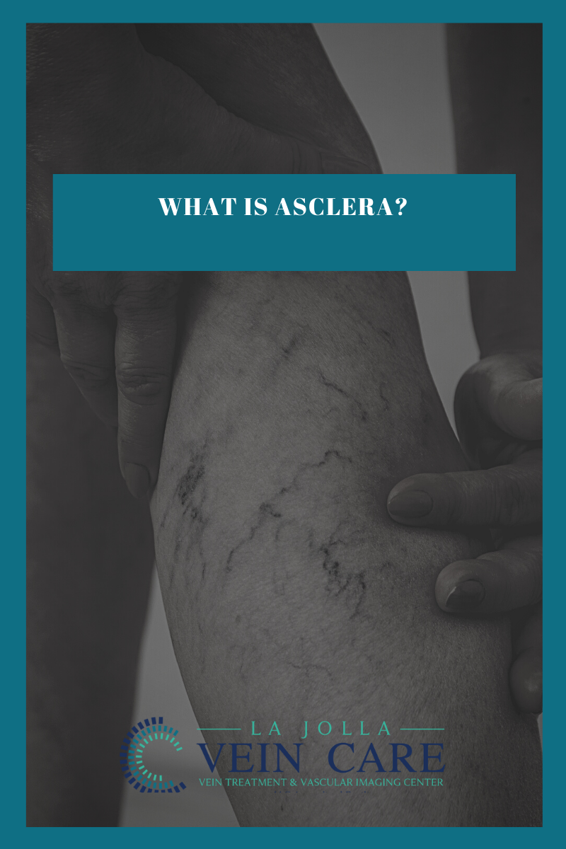
A variety of sclerosant medications can be used for veins, but at La Jolla Vein Care, we prefer polidocanol (brand name Asclera), because it is the most comfortable for the patient and has the lowest risk of side effects. It is a detergent-type medication that injures the inner lining of the veins. This causes the vein to stop flowing, then eventually close down completely. Polidocanol has been well-studied and has been approved by the FDA since 2010 for the treatment of varicose veins and has been used in Europe for decades. Polidocanol is turned into a foam to treat larger veins. This is an off-label use of the sclerosant but very effective and well tolerated. Asclera (polidocanol) Injection is a prescription medicine that is used in a procedure called sclerotherapy to remove unwanted veins on your legs. It is administered by a healthcare provider to treat two types of veins:


  • Uncomplicated spider veins (very small varicose veins ≤ 1 mm in diameter)
  • Uncomplicated small varicose veins (1 to 3 mm in diameter) known as reticular veins

Asclera has not been studied in varicose veins more than 3 mm in diameter. La Jolla Vein Care providers have extensive experience with foam sclerotherapy. 


ADVERSE REACTIONS: 


  • injection site hematoma
  • injection site irritation 
  • injection site discoloration 
  • injection site pain 
  • injection site itching 
  • injection site warmth 
  • neovascularization 
  • injection site clotting


How often do I need treatment to see results?

The number and frequency of treatments depends on the size of the vessels and their location. Ask your provider about an individualized treatment approach that is right for you.

How does Asclera work?

Asclera is a sclerosing agent that is injected into the vein. It works by damaging the endothelium, the inside lining of blood vessels. This causes blood platelets and cellular debris to attach to the lining of the vessels. Eventually, cellular debris and platelets cause the blood vessel to clot. Over time, the clotted vein will be replaced with tissue.

How satisfied are patients who use Asclera?

88% of clinical study patients were satisfied or very satisfied with their Asclera treatment after 12 weeks.

How long is each Asclera session?

A typical session lasts 15 to 45 minutes. Generally, 1 to 3 injections may be necessary to treat a given spider or reticular vein. Repeat treatment sessions may be necessary. Any additional treatment sessions are usually separated by 1 to 2 weeks.

What should I expect after being treated with Asclera?

Following treatment, you’ll need to wear compression stockings day and night for 2 to 3 days, then for 2 to 3 weeks during the daytime, as directed by your provider. Compression stockings are designed to apply pressure to your lower legs, helping to maintain blood flow, and reduce discomfort and swelling. Compression helps your legs heal and is necessary to reduce the risk of deep vein thrombosis.


Are there activities I should avoid after receiving an Asclera Injection?

For 2 to 3 days following the treatment, avoid:

  • Heavy exercise
  • Sunbathing
  • Long plane flights
  • Hot baths, hot tubs, or saunas

Possible side effects to Asclera treatments:

Temporary side effects may occur at the site of the injection, including:

  • Bruising
  • Raised red areas
  • Small skin sores
  • Darkened skin in the form of lines or spots
  • Multiple tiny red blood vessels

These side effects usually go away within a few days to several weeks. Some side effects may take months or years to resolve.

For more information or to book an appointment for Asclera therapy, please call our office at 858-550-0330.

You can visit the Asclera website here.


What is Asclera?2022-12-13T13:07:15-08:00

23, 3, 2022

What is Vascular Disease?

2022-03-23T11:47:16-07:00

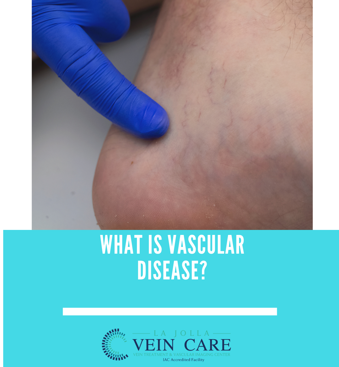
What is Vascular Disease? 

Your body contains an amazing, intricate system of arteries and veins. Arteries bring oxygen-rich blood from the heart to every inch of the body; veins return the blood back to the heart and lungs for more oxygen.

Most people know that heart disease can occur when blood vessels become clogged with plaque and cholesterol, but many are unaware that the same problem, often called hardening of the arteries (or atherosclerosis), can occur in any artery of the body.

Venous problems occur in the veins, often resulting in leg swelling, deep vein thrombosis (blood clots) and chronic leg problems.

Common types of artery disease include:


Carotid artery disease and stroke

Carotid arteries in the neck bring oxygen to the brain. Patients who have had a stroke, a transient ischemic attack

(TIA) or have been told they have an abnormal sound in that artery, will be referred to a vascular surgeon for diagnosis and possible treatment.

Aneurysms

Aneurysms are bulges that develop in a weakened part of an artery. While these can occur in the brain, they are most common in the aorta, which is the body’s biggest artery, running from the heart through the abdomen. Aneurysms can also occur in the pelvis or the knee. They can enlarge when the heartbeat pushes against the weakened wall. In the aorta and pelvis, ruptured aortas can be fatal.

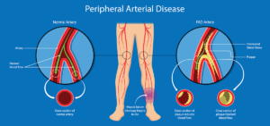
Peripheral arterial disease (PAD)

Patients with hardening of the arteries start to lose blood flow to the limbs. Early on, this may cause pain when walking, but as the disease progresses, it can cause painful foot ulcers, infections, and even gangrene, which could require amputation. People with PAD are three times more likely to die of heart attacks or strokes than those without PAD.

Common Vascular Disease Problems: 


  • Varicose veins which are often cosmetic, but can also be a sign of a more serious venous disease.
  • Chronic venous insufficiency is a potentially painful condition that is caused by blood reflux or clotting.
  • Deep vein thrombosis a blood clot in a deep leg vein.


Who are Vascular Surgeons:


Vascular surgeons train in vascular disease for five to seven years after medical school. Board certified vascular surgeons can treat any vascular condition with any treatment—medication, minimally invasive endovascular surgery, or open surgery. Many vascular conditions are lifelong, and your vascular surgeon may become a lifelong care partner. Be sure to ask to be referred to a board certified vascular surgeon.


What is Vascular Disease?2022-03-23T11:47:16-07:00

The Best Way to Treat Reticular Veins

2022-03-17T10:17:12-07:00

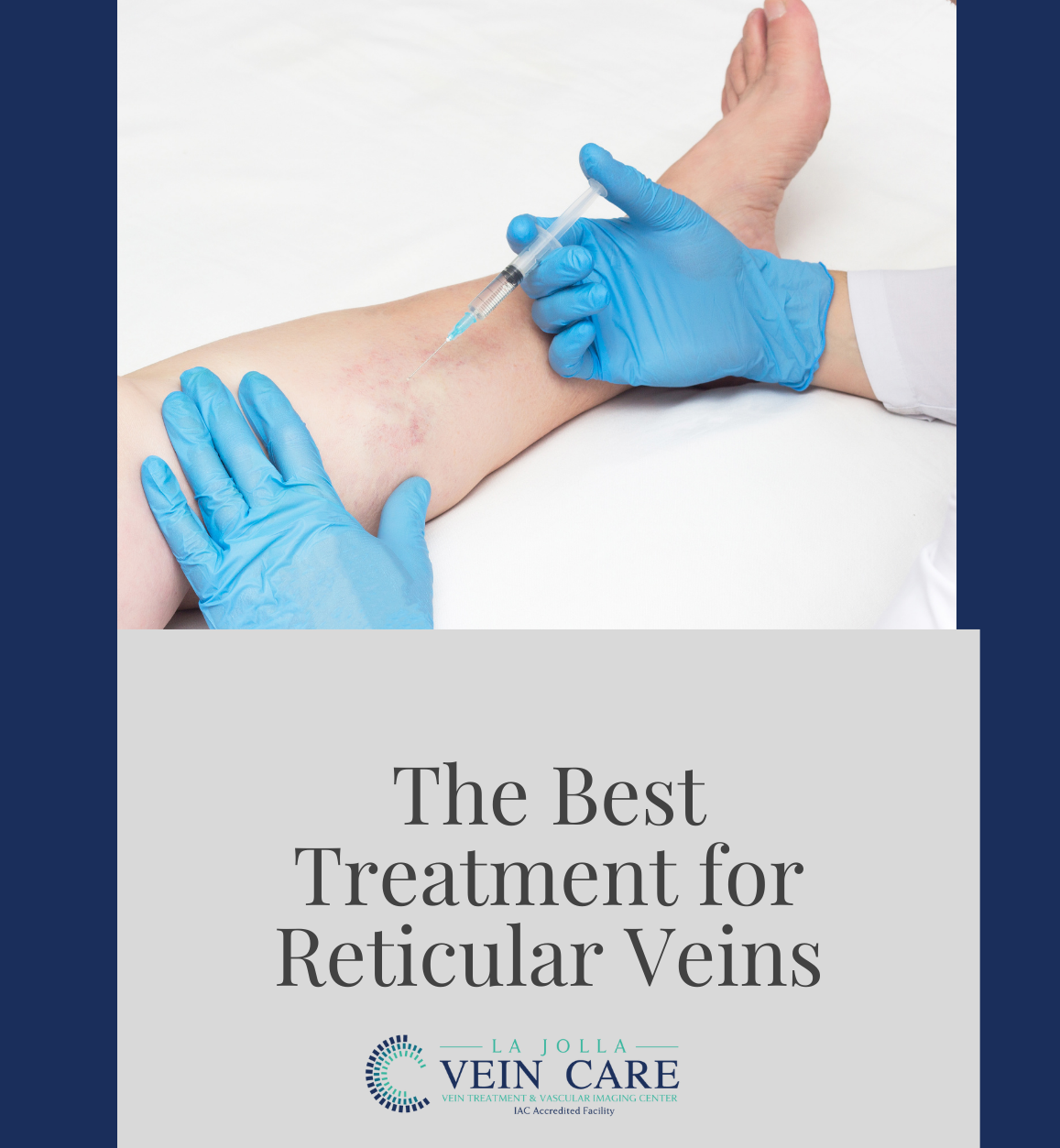
The best treatment for Reticular Veins is through a painless alternative to traditional sclerotherapy: Cryo-Sclerotherapy.

Why have I been offered cryo-sclerotherapy?


We recommend patients undergo cry-sclerotherapy to mainly treat spider veins and small varicose veins at the skin surface (also known as reticular veins). Traditional sclerotherapy involves injection of a medication into the vein using a tiny needle. This causes the vein to collapse and eventually fade away. The medication will be administered in a liquid and /or foam formulation, depending on the size of the veins. Cryo-sclerotherapy is the combination of traditional sclerotherapy in combination with the use of a cryo device. The cryo device delivers puffs of cold air simultaneously during sclerotherapy. The result  is the cold air anesthetizes the skin so the treatment is relatively painless. This is especially helpful for treatment of sensitive areas like the inner ankles and thighs. Instead of feeling the needle prick, you will feel puffs of cold air instead. The cold air may also reduce bruising and pain. 


What should I expect on my treatment days? 


You will sign your consent form then change into shorts provided by the office. We will clean your skin with alcohol. The sclerosant medication will then be injected into your veins with a fine needle. Just before the needle is used, you will feel puffs of cold air. The cold air will anesthetize the skin, so that the prick from the needle is not felt or lessened. Every time the needle is used, you will feel puffs of cold air instead. After your treatment, we will help you into your compression stockings, then you will walk for 30 minutes prior to getting in your car. It is normal for your legs to be achy and tender to the touch after treatment. 


What should I do after treatment?


You will be instructed on how long to wear compression stockings after treatment, depending on the size of the veins that are treated. Most patients wear thigh-high compression stockings continuously for 1-3 days, then another 7 days. 


You may shower with the stockings on or take a quick cool shower with them off. 


You should walk 30 minutes twice daily after treatment and move your legs frequently throughout the day with short walks and/or calf exercises. This avoids pooling of blood in the legs. Avoid prolonged sitting during the day. 


It is normal to have aching in the treated veins. This responds well to walking, ice packs, and anti-inflammatory medications such as ibuprofen (Advil, Motrin) and naproxen (Aleve). 


What should I avoid after treatment? 


For at least two weeks after treatment, you should avoid strenuous exercise (anything more than a brisk walk), heavy lifting, saunas or hot tubs, and leg massages. All of these dilate the superficial veins and interfere with their healing. You should also avoid airline travel for two weeks after treatment. 


What are the possible side effects? 


You will likely have some bruising over the injection sites, then the treated veins may become firm and blue. 


The skin overlying treated veins may also develop brownish hyperpigmentation as the blood products within those veins are absorbed by the body; in some patients it can take up to a year to fade. Some patients temporarily develop some very fine, pink spider veins in areas where veins have been treated (telangiectatic matting). These usually resolve spontaneously over several weeks but occasionally require additional treatment to clear. 


Many patients will feel their veins look a bit worse in the first month after sclerotherapy, but through the best treatment for reticular veins, cryo-sclerotherapy, patients have reported it to be a more painless approach for treatment.  


The Best Way to Treat Reticular Veins2022-03-17T10:17:12-07:00
Go to Top