15, 7, 2022

Vein Health & Your Nutrition

2022-06-25T17:32:08-07:00

One’s vein health and nutrition is very important. One contributing factor that can influence your vein health is through your diet and what foods you consume on a daily basis. There are nutrient dense foods that help contribute to your health. For vein health specifically, foods that are rich in flavonoids may improve symptoms of venous disease. Flavonoids help protect plants from environmental toxins and help repair damage. They can be found in a variety of foods, such as fruits and vegetables. When we eat foods rich in flavonoids, it appears that we also benefit from this “antioxidant” power. It’s important to include in your daily nutrition foods that have antioxidants. 


In addition to having antioxidant qualities, research shows that flavonoids have other potential influences on vascular health, such as lowering blood pressure, improving blood flow to the brain and heart, and making blood platelets less sticky and able to clot.Flavonoids are also well known for their ‘venoactive’ effects on the blood vessels and have been proven to reduce symptoms of venous disease such as leg aching, heaviness and swelling.

Foods that are flavonoid rich include cocoa and chocolate, bell peppers, spinach, broccoli, grapes, blueberries, cranberries, apples, peanuts, onions, garlic, tea and red wine.

Besides improving your nutrition for your vein health, remind the cook to use compression socks- long hours of standing in the kitchen can cause leg fatigue, heaviness and swelling and eventually be a sign of venous disease. 

If you experience any vein disease symptoms, please call our office at (858)-434-5998 to schedule a consultation with one of our knowledgeable doctors at La Jolla Vein and Vascular. 


For more information on vein health please check out our Youtube Channel or visit our helpful guide of resources


Vein Health & Your Nutrition2022-06-25T17:32:08-07:00

Before & After Spider Vein Treatment

2022-06-25T16:09:12-07:00

What are spider veins?

Spider veins are the fine, thread-like reddish veins at the surface of the skin. These veins are not healthy. Veins are often considered a cosmetic issue, but they can be associated with underlying feeder veins, not visible to the naked eye.  Feeder veins are the ‘blue veins’ also known as ‘reticular veins,’ that give rise to spider veins. Spider veins may also be a sign of underlying venous insufficiency. For example, spider veins located in the inner ankles and inner thighs can indicate an underlying problem with the saphenous vein. As a result, an ultrasound examination may be recommended to identify and effectively treat the underlying source of these veins.

What are the symptoms?

Vein conditions affect people differently. For some, these veins can be a painless cosmetic concern, and for others they may cause symptoms. The most common symptoms of spider veins are burning, throbbing, and localized pain. They can also feel hot and itchy and bleed.

What causes them?

Spider veins in the legs are caused by the same condition that causes varicose veins. Leaky vein valves allow blood to pool within the veins causing them to stretch and become enlarged. These veins on other areas of the body, such as the face, and chest can be caused by sun damage, hormone changes or liver disease. Hormones, such as with pregnancy, birth control or hormone replacement therapy can weaken the vein wall.

Below you can see the transformation of 3 of our patients who received vein treatment.

spider veinsspider veinsspider veins

If you experience any vein disease symptoms, please call our office at (858)-434-5998 to schedule a consultation with one of our knowledgeable doctors at La Jolla Vein and Vascular.

For more information on vein health please check out our Youtube Channel or visit our helpful guide of resources.

Before & After Spider Vein Treatment2022-06-25T16:09:12-07:00

Vein Health & Working From Home

2022-12-13T13:18:14-08:00

Prolonged desk work can increase the risk of blood clots in leg veins and decline your vein health.

As many Americans are working from home, we have received many calls from patients regarding thrombophlebitis, which is a big concern for vein health. This is not a new scenario for us, over the years frequently patients come in for assessment of leg pain after working on their computers for 12 hours straight.

ThromboPhlebitis refers to blood clots in the leg veins and inflammation of the surrounding vein. The inflammation causes leg pain and redness of the surrounding skin. You may also feel hard lumps in the leg veins.

Inactivity, sitting for long periods of time, like sitting at your desk, working on your computer for many hours without walking or getting up for a light walk or exercise, affects the blood flow in the leg veins. Your calf muscle pump, which is used when walking and moving about helps to move blood back to your heart. Sitting for too long of periods, increases the risk of your blood pooling in the leg veins and can even form blood clots. Blood clots that form in the deep veins are serious as they can travel to the heart and lungs (deep venous thrombosis). Blood clots that form in the surface veins, are called superficial thrombophlebitis (STP).  With STP, you can typically feel lumps that can be painful. People who have varicose veins, have an increased risk of developing STP especially with inactivity. Being mindful about your vein health while working remotely will help in the longer run when it comes to vein disease. 

Tips to Avoid Phlebitis When Working From Home:

  1. Avoid sitting at your desk or computer for longer than 45 minutes without taking a break to get up and walk about.
  2. Try to walk at least 30 minutes a day or other exercise
  3. When sitting, do calf raises to pump the calf veins.
  4. If you have varicose veins, you should wear compression stockings during the day time.

If you experience any vein disease symptoms, please call our office at (858)-550-0330 to schedule a consultation with one of our knowledgeable doctors at La Jolla Vein and Vascular. 


For more information on vein health please check out our Youtube Channel or visit our helpful guide of resources


Vein Health & Working From Home2022-12-13T13:18:14-08:00

Spider Veins: Before & After Transformation

2022-06-25T16:03:26-07:00

What are spider veins?

Spider veins are the fine, thread-like reddish veins at the surface of the skin. These veins are not healthy. Veins are often considered a cosmetic issue, but they can be associated with underlying feeder veins, not visible to the naked eye.  Feeder veins are the ‘blue veins’ also known as ‘reticular veins,’ that give rise to spider veins. Spider veins may also be a sign of underlying venous insufficiency. For example, spider veins located in the inner ankles and inner thighs can indicate an underlying problem with the saphenous vein. As a result, an ultrasound examination may be recommended to identify and effectively treat the underlying source of these veins.

What are the symptoms?

Vein conditions affect people differently. For some, these veins can be a painless cosmetic concern, and for others they may cause symptoms. The most common symptoms of spider veins are burning, throbbing, and localized pain. They can also feel hot and itchy and bleed.

What causes them?

Spider veins in the legs are caused by the same condition that causes varicose veins. Leaky vein valves allow blood to pool within the veins causing them to stretch and become enlarged. These veins on other areas of the body, such as the face, and chest can be caused by sun damage, hormone changes or liver disease. Hormones, such as with pregnancy, birth control or hormone replacement therapy can weaken the vein wall.

Below you can see the transformation of 3 of our patients who received vein treatment.

spider veinsspider veinsspider veins

If you experience any vein disease symptoms, please call our office at (858)-434-5998 to schedule a consultation with one of our knowledgeable doctors at La Jolla Vein and Vascular.

For more information on vein health please check out our Youtube Channel or visit our helpful guide of resources. 

Spider Veins: Before & After Transformation2022-06-25T16:03:26-07:00

Vein Health: Helpful Anatomy Phrases to Better Understand

2022-12-13T14:15:11-08:00

If you’re not a vascular specialist, it may be difficult to understand the words used to describe your vein condition and understand vein health. Vein terms are not used in everyday life.  Especially, when it comes to the anatomy of the veins. Below is a glossary of the terms, we as vein specialists, use on a daily basis but may be foreign to the patient.  This is provided from the American Vein and Lymphatic Society (AVLS) healthyveins.org.

Anatomical Vein Terms for Vein Health:

Anterior: The front of a body part.

Posterior: Back of a body part.

​Deep Leg Vein: The veins that are deep in the thigh and leg that carry blood back to the heart.

​Connective Tissue: This strong tissue is the white tissue that covers muscle.

Great Saphenous Vein: (sometimes called GSV or long saphenous vein) A long vein that can be seen just in front of the ankle bone. This vein travels along the inside of the leg and thigh (about one-half inch beneath the skin in the thigh) until it empties into the deep vein called the common femoral vein in the groin.

Perforating Vein: A vein that passes directly from a superficial vein to a deep vein.

Popliteal Vein: The deep vein located behind the knee. The small saphenous vein originates from the popliteal vein.

Small Saphenous Vein: (sometimes called SSV, Lesser Saphenous Vein, or LSV) A superficial vein that starts at the outside of the foot and travels up the back of the calf where it empties into the deep vein (popliteal vein) in the crease of the knee.

​Spider Vein: A tiny varicose vein that may be blue or red color that does not protrude above the skin surface and frequently looks like a spider. Common medical terms you may hear for spider veins are telangiectasias and telangiectatic veins. Slightly larger varicose veins that do not protrude above the skin are called reticular veins.

​Superficial Vein: Any vein in the lower extremity above the deep fascia that covers the muscles of the thigh and leg.

​Varicose Vein: A bulging vein that protrudes past the skin surface and usually measures greater than one-fourth of an inch (6.4 mm) in diameter.

Vein Function Terms

Competent Vein: Blood flow occurs in the proper direction back to the heart. Also referred to as Having No Reflux Or Normal Flow Direction.

Incompetent Vein: Blood flows in the wrong direction. Also referred to as a vein that has reflux.

Reflux: Blood that flows backward in the veins.


Anatomical Images and Explanations

Normal Blood Flow: After the blood has been replenished with oxygen in the lungs, it is pumped to the body by the heart. Blood that is pumped to the lower extremities is pumped back to the heart partially by the action of the calf muscle pump. The blood returning from the lower extremities in the deep and superficial veins goes past a series of one-way valves. These valves stop the blood from flowing backwards in the vein.

Reflux: The valves in the veins close just as blood begins to flow backwards. If the valves do not close properly, the blood falls backwards through the poorly closing or leaking valves. The veins downstream that are now unprotected by valves further upstream are exposed to the weight of an increasingly high column of blood. These downstream veins cannot endure the pressure of the column of blood and expand becoming snake-like in appearance. This causes the vein to bulge through the skin surface and become varicose veins.

If you experience any vein disease symptoms, please call our office at (858)-550-0330 to schedule a consultation with one of our knowledgeable doctors at La Jolla Vein and Vascular.

For more information on vein health please check out our Youtube Channel or visit our helpful guide of resources. 

Vein Health: Helpful Anatomy Phrases to Better Understand2022-12-13T14:15:11-08:00

20, 6, 2022

Pelvic Venous Congestion Treatment

2022-05-23T15:15:40-07:00

Pelvic Venous Congestion (PVCS) Treatment

What are the treatment options for Pelvic Venous Congestion (PVCs)? Available treatment options for PVCs include pain-relieving progestin hormone drugs, ovarian function-blocking Gonadotropin-releasing hormones, vein embolization, and surgery to rid of damaged veins, uterus, and ovaries. Customarily, your doctor will start you on the medications first to relieve pain. If the medications fail, then they may advise on the next step, usually the invasive options. Vein embolization is the most commonly used procedure. Here’s a look at what it involves:

-Pelvic Venous Embolization

-Sclerotherapy

-Radiofrequency Ablation (RFA)

pelvic venous congestion


Pelvic Venous Embolization

Treating PVCS diseases like pelvic and labial varicose veins requires resolving the backward flow of blood in the ovarian and pelvic veins through embolization. Embolization is an outpatient procedure that uses catheter technology to access the venous system to the problematic ovarian veins. The procedure is painless and requires no major incisions. The physician embolizes each problem vein (blocking blood flow) with vascular plugs or coils. Doing so permanently seals off the varicose veins, diverting blood flow up through healthy veins. The physician will also deliver sclerosing medication to lower the pressure in the pelvic veins and relieve the pain. Once blood can flow smoothly through healthy veins in the pelvic and labial area, the pain disappears. Patients can usually return to work and light activities the day following the procedure and see a full recovery in about one week; at least 7 in 10 women who undergo the procedure report feeling better. This procedure is usually performed by a trained interventional radiologist in the X-ray department.


Sclerotherapy

Sclerotherapy is a minimally invasive procedure that injects sclerosing solutions into the veins. It is used to treat spider, reticular and varicose veins. It also blocks the unsightly or non-functioning veins to gradually improve appearance. This generally involves multiple treatments over the course of several months for pelvic venous congestion. 

Radiofrequency Ablation is a minimally invasive, image-guided procedure used to treat varicose veins. An RFA utilizes high-frequency radio waves directed through a thin tube to create intense heat within the varicose vein. This closes up the problem vessel so blood cannot flow through it, redirecting blood flow into healthy veins to allow the blood to return to the heart. Eliminating the unhealthy vein reduces the vein bulging and minimizes leg pain and swelling. It is performed as an outpatient procedure with minimal recovery and discomfort.


When Should I Contact My Doctor?

Some women have enlarged veins with no symptoms; others have enlarged veins that cause pains and aches. In most cases, these don’t require a medical emergency. The symptoms should reduce as you head into menopause. However, in case of chronic, unbearable pain, nothing is stopping you from seeking medical help. In fact, see your healthcare giver right away for help.

To get the most out of the pelvic venous congestion consultation, here’s what to do:

-Know the purpose of the visit and what you want to get from it

-Before visiting the doctor, put down questions you need them to answer.

-Bring company along to help ask questions and note down points from the doctor.

-Write down the name of the diagnosis, its tests, and treatment options explained by the doctor. -Also, take notes of any new instructions during each visit.

-Know the reasons for new medication prescriptions, their benefits, and their risks.

-Inquire whether it’s possible to treat your condition another way

-Know why and what the results of a test procedure mean

-Ask what will happen if you don’t undergo testing or take medication.

-Ask whether a follow-up appointment is available and note down the visit’s time, date, and reason.

-Ask the doctor for their contacts in case you need a further chat.


 For more information on pelvic venous congestion and to book a consult, please give our office a call at 858-550-0330.

For more information please check out our Youtube Channel at this link.

Pelvic Venous Congestion Treatment2022-05-23T15:15:40-07:00

Arterial Treatments

2022-05-23T14:56:03-07:00

Arterial Treatments


Peripheral Arterial Disease – Treatment

Arterial treatments for the peripheral arterial disease (PAD) has two main goals. One is to manage the symptoms, including leg pain, allowing you to resume your normal physical activities. The second one is to top the progression of atherosclerosis in the body, which reduces the risk of stroke and heart attack.

The good news is that through early intervention, you can achieve these goals through lifestyle changes. Quitting smoking is one of the most significant things you can do to stop the progression of Pad. Walking and exercising regularly following supervised exercise training can improve your symptoms. However, if your peripheral artery disease is at an advanced stage, you will need additional medical treatment. Here are some of the standard peripheral artery disease treatment options available.

Atherectomy

A lower extremity atherectomy is a minimally invasive surgical procedure involving a catheter to remove plaque from arteries. The doctor usually makes a small incision in the artery to insert the catheter. The procedure is performed under local anesthesia to prevent pain. A catheter collects removed plaque in a chamber placed at its tip, ensuring that all the particles are removed from the artery. Depending on the amount of plaque in your arteries, your doctor will repeat the procedure several times to ensure optimal blood flow.

Why Do I Need This Procedure?

The primary goal for the atherectomy is to eliminate plaque buildup in the artery. You may need this procedure when your arteries become too narrowed or blocked due to increased plaque. When the arteries are blocked, blood flow is restricted, meaning that your tissues will not have sufficient oxygen and nourishment they need to survive. Insufficient blood flow causes the muscle in your feet to cramp and lose strength. The procedure is not ideal for all people. Before performing the procedure, the doctors customize to meet the patient’s unique needs. 

Where is the Procedure Performed?

We usually perform the procedure in our facility. Our board of certified doctors have undergone rigorous training to ensure the best possible results. We also have a state-of-the-art facility with modern atherectomy tools that ensure success even for complex conditions. Our doctors also have a vast wealth of experience in this field. Having performed thousands of atherectomy procedures is sure that you are in the right hands. 

How do I Prepare for an Atherectomy?

A few days before the procedure, the doctor usually performs a pre-procedure test to ensure that it is safe for you to undergo the procedure. The doctor may recommend discontinuation of using certain medications days before the procedure. Our healthcare team will also provide you with specific instructions to prepare for the procedure.


Arterial Treatments2022-05-23T14:56:03-07:00

Causes of Osteoarthritis

2022-05-13T17:16:22-07:00

Causes Of Osteoarthritis (OA)


Although Knee Osteoarthritis affects as many as 45 percent of people in their lifetime, the exact cause is unknown. However, medical practitioners have discovered several risks that increase the chances of developing knee osteoarthritis. These include:

Advanced Age

Obesity

Joint Trauma

Family History

Congenital Disabilities or Illnesses

Chronic Injury and Joint Stress

Lack of Exercise

Poor Muscle Tone

Female Sex


Advanced Age

Your susceptibility to developing osteoarthritis increases with age. As you grow old, the cartilage in the knee joint wears down, making it prone to damage and thinning. According to research done by the Arthritis Foundation, about 19 percent of people over 45 years have knee OA. The research further indicates that 37 percent of people over 60 years have the condition, although not all will have the symptoms.


Obesity

The knees support the weight from the thighs upwards. Obese people are at two times more risk of developing knee arthritis than individuals who are not.


Joint Trauma

Surgery, a broken bone, or severe injuries that affect the knee joint can also lead to knee osteoarthritis. These symptoms may not be present immediately after the injuries but may reveal years later.


Family History

Hereditary genes also play a crucial role in the susceptibility of knee arthritis. For instance, if your mother had knee OA, you are more likely to develop the condition than people with no family history of the condition are.


Congenital Disabilities or Illnesses

Septic arthritis, gout, poor bone alignment, congenital conditions, and metabolic disorder can also increase your susceptibility to contracting knee arthritis.


Chronic Injury and Joint Stress

Individuals whose daily activities involve heavy lifting while standing, crawling, or squatting tend to experience mini-traumas. These mini traumas can lead to knee osteoarthritis over time—bodybuilders and other athletes who participate in high-impact sports are prone to knee osteoarthritis development.


Lack of Exercise

Although putting too much stress on your knee joint can lead to arthritis, lack of exercise can also cause OA. Exercise helps in boosting the cartilage’s health and repair. Additionally, the knee joints contain fluid-rich nutrients that need to be circulated in the knee joint capsule. Regular exercise promotes proper circulation of the joint fluid.


Poor Muscle Tone

Poor hamstring, calf, and quadriceps muscle development can put more stress on the knee’s bones and cartilage, causing knee osteoarthritis.


Female Sex

Research shows that women are about 40 percent more likely to develop knee arthritis than men are.


 So, if this is you and you’ve been experiencing pain, please give our office a call at 858-550-0330. 


For more information please check out our Youtube Channel at this link. 


Causes of Osteoarthritis2022-05-13T17:16:22-07:00

Signs of Osteoarthritis

2022-05-30T13:29:11-07:00

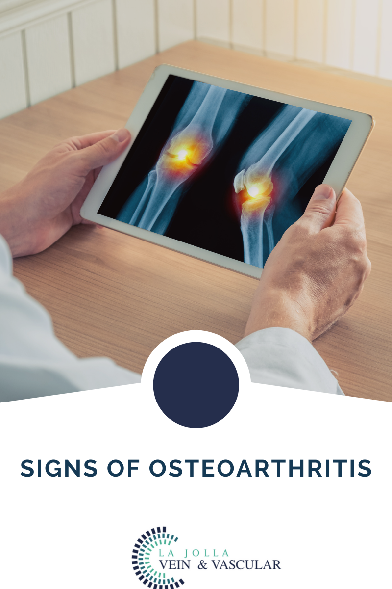
For most people, the knee osteoarthritis symptoms come and go, worsening over time. While it is easy to dismiss early knee OA signs and symptoms, the symptoms become worse if left untreated, causing mobility issues. 


Some of the common OA signs and symptoms include:


Knee Pain: Knee pain is the most reported OA symptom. The pain description often varies from one person to the other depending on their condition and situation. Sometimes the pain may come and go, while the person may experience chronic low levels of pain with intermittent intense pain flare-ups. On the other hand, you may experience a dull, aching. Most often, knee OA pain gets worse, especially when doing activities that put a strain on the knee, such as walking upstairs or squatting.


Swelling: Damaged cartilage causes the femur and tibia bones to rub together, leading to irritation. The irritation forces the body to produce excess fluid at the joint, making the knee swell.


Stiffness: The swelling and friction in the knee make the joint stiff. You may often experience 30 minutes of stiffness after sitting for an extended period or early in the morning. Knee stiffness can also be accompanied by reduced motion range and visible swelling.


Redness and Warmth: The irritation develops when the knee joint’s bones rub each other causing the skin over the knee to become warm and red. These signs could also indicate an infection that requires medical attention.


Reduced Range of Motion: The swelling in the knee limits your range of motion. Mild to moderate knee OA makes it hard for you to bend or straighten the knee completely.

Worsening Symptoms with Inactivity

Your knees tend to become stiff after long periods of sitting or sleeping. In most cases, you will notice pain and stiffness after waking up in the morning or sitting for long hours.


Popping or Crunching: A popping or crunching sound when bending your knee indicates knee joint friction.


Buckling or Locking Up: People with moderate to severe knee OA can also cause a sensation of knee-buckling. The sensation develops when there are uneven flaps or grooves, or cartilage breaks off and sticks in the joint.


 So, if this is you and you’ve been experiencing  osteoarthritis pain, please give our office a call at 858-550-0330. 


For more information please check out our Youtube Channel at this link. 


Signs of Osteoarthritis2022-05-30T13:29:11-07:00

27, 5, 2022

What causes uterine fibroids?

2022-04-29T17:20:54-07:00

What Causes Uterine Fibroids?

While the science is not completely settled on what exactly causes or is behind the formation of uterine fibroids in human beings, there are certain factors that researchers believe play significant roles in triggering their growth or development.

Genetics

In 50 percent of cases, patients will be found to have some genetic abnormality. In many instances, some erroneous translocation will be discovered on some chromosomes.

Hormones

While we may not be completely certain as to what lies at the root cause of uterine fibroids, it has been clearly demonstrated that hormones play a large role in determining their growth or progression. While a woman is pregnant, they will see rapid growth due to the presence of the hormones estrogen and progesterone. Should the patient use anti-hormone medication, the opposite will happen – the fibroids will shrink. Furthermore, fibroids will stop growing or begin shrinking once a patient reaches menopause, which is the point at which these hormones also cease being produced in the female body.

uterine fibroids

Ultrasound image of lower abdomen, ovary and uterus with tumor or uterine fibroid, leiomyoma of female woman patient for gynecological medical exam, analysis and test

Testing and Diagnosis of Uterine Fibroids

In many cases, especially those whereby uterine fibroids do not present any noticeable symptoms for the patient, this condition will be discovered in a regular pelvic exam. This is a routine checkup carried out by gynecologists. While this checkup might determine the presence of a mass or lump on the uterus, further testing will have to be carried out to make a definite diagnosis.

These testing methods include:

Note: Upon learning that fibroids are a type of tumor, the foremost concern for many patients is whether the tumor is cancerous or might predispose them in some way to developing cancer. Fibroids are rarely cancerous, with the chances of such fibroids (leiomyosarcomas) arising at less than one in a thousand.  Additionally, the presence of fibroids in no way increases a patient’s chances of developing any type of cancer.


What causes uterine fibroids?2022-04-29T17:20:54-07:00
Go to Top