6, 6, 2022

What are Pelvic & Labial Varicose Veins

2022-05-12T12:46:22-07:00

Pelvic & Labial Varicose Veins

labialvaricosities

Labial varicosities are a common vascular condition that is now treated at La Jolla Vein and Vascular. Most people understand that varicose veins can be found in the legs and feet. And while this is mostly true, they can also manifest in the vulva. This is generally why most women who develop from vulva varicosities don’t notice it until the condition begins to worsen.


What are Labial Varicosities?


Vulvar varicosities, also commonly referred to as labial varicosities, are a condition when the veins in the labia minora or Majora become dilated.


Like any other part of the human body, the vulva is interconnected through an extensive reach of large and small blood vessels. During pregnancy, the increase in weight and pressure may reduce blood flow to the lower body and genitals. This will then cascade into the condition known as Labial varicosities.


While no one is fully exempt from developing vulvar varicose veins, the condition is more apparent in pregnant women. Moreover, pregnant women are more susceptible to developing pelvic varicosities due to the pressure and weight exerted by the baby.


However, in most cases, the condition does go away after giving birth. Ideally, you should start feeling better within six to eight weeks from the delivery time. Unfortunately, in some women, the dilated veins do not recover independently and need medical intervention to help fix the condition and get you back to your normal self from labial varicose veins. 


Most people understand that varicose veins can be found in the legs and feet. And while this is mostly true, they can also manifest in the vulva. This is generally why most women who develop from vulva varicosities don’t notice it until the condition begins to worsen.

There are numerous veins supplying blood to and from the vulva. And since they all differ in size, there’s no sure way to tell if you’re developing pelvic varicosities. The impact may leave them moderately swollen from the varying size of the veins, especially in the smaller veins. Larger veins may get twisted and become painful over time. This further goes to assert the importance of consulting a healthcare professional if you’re uncertain.


 So, if this is you and you’ve been experiencing pain or any other symptoms, please give our office a call at 858-550-0330. 


For more information please check out our Youtube Channel at this link. 


What are Pelvic & Labial Varicose Veins2022-05-12T12:46:22-07:00

Symptoms of Pelvic Venous Congestion

2022-05-12T10:49:38-07:00

Symptoms to look for in Chronic Pelvic Venous Congestion


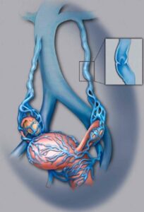
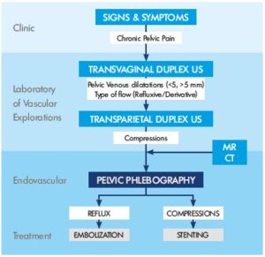
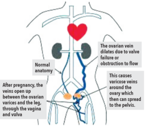
Pelvic Venous Congestion also known as (PVCS) is a common venous disease that La Jolla Vein and Vascular is now treating. Most people know varicose veins mostly affect the feet and legs. But varicose veins can also occur in the pelvis causing chronic pelvic pain in women; this condition is known as Pelvic Venous Congestion Syndrome (PVCs) or ovarian vein reflux.

pelvicvenouscongestion

PVC lasts longer than half a year. PVCs are commonly experienced for the first time during or following pregnancy. It is characterized by a heavy aching feeling that may get worse as pregnancy progresses. In most cases, you feel the pain on the left side only. But at other times, you may feel the pain on both the left and right sides. PVCs pain is usually worse during the evenings.

Certain factors also aggravate pelvic venous congestion pain; these include:

  • When you change posture

  • Sex

  • Standing for extended periods

  • Walking

  • Menstrual period

Apart from pelvis pain, other pelvic venous congestion symptoms include:

  • Pain during or after intercourse

  • Sudden urge to urinate

  • Lower back pain

  • Irritable bowel syndrome (diarrhea and constant abdominal pain accompanied with constipation)

  • Deep dyspareunia – pain during intercourse

  • Feeling one’s legs fuller

  • Engorged and distorted veins around the vagina, vulva, inner thigh, buttocks, and sometimes down the legs

pelvic venous congestion

 So, if this is you and you’ve been experiencing pain, please give our office a call at 858-550-0330.


For more information please check out our Youtube Channel at this link.


Symptoms of Pelvic Venous Congestion2022-05-12T10:49:38-07:00

What is Pelvic Venous Congestion?

2022-05-12T10:42:52-07:00

Pelvic Venous Congestion (PVCS)


Pelvic Venous Congestion also known as (PVCS) is a common venous disease that La Jolla Vein and Vascular is now treating. Most people know varicose veins mostly affect the feet and legs. But varicose veins can also occur in the pelvis causing chronic pelvic pain in women; this condition is known as (PVCs) or ovarian vein reflux. Herein is a look at the condition, the risk factors, causes, symptoms, and treatment options. Read on to find out more.

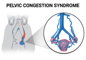
What is pelvic venous congestion syndrome (PVCs)?

pelviccongestionsyndrome

Pelvic Venous congestion is the enlargement of blood vessels in the pelvis due to faulty vein valves in the lower abdomen. See, veins have valves to guide blood flow towards the heart. However, these valves become faulty in some cases due to damage or other problems causing blood to flow backward. When this happens in the lower abdomen, blood builds up in the area, causing the veins to enlarge and change shape.

pelvic venous congestion

PVCs is when varicose veins are in your pelvis. This engorgement or congestion of blood vessels in the lower abdomen can cause chronic, and unbearable pain. It affects at least 1 in three women at some point during their lifetime. Chronic pelvic venous congestion syndrome may last longer than six months. It is not associated with period pain at all. PVCs are common among women who have given birth more than once before. So, if this is you and you’ve been experiencing pain, please give our office a call at 858-550-0330. 

For more information please check out our Youtube Channel at this link. 


What is Pelvic Venous Congestion?2022-05-12T10:42:52-07:00

Uterine Artery Embolization (UFE) vs. Hysterectomy

2022-04-29T17:33:47-07:00

Benefits of UFE Over Hysterectomy

UFE ( Uterine Artery Embolization ) is a minimally invasive procedure that is effective for treating uterine fibroids and is considered an alternative to uterine fibroid surgery removal. UFE is typically performed as an outpatient procedure, which means that most women go home the same day for recovery.

UFE has several benefits over hysterectomy. Besides involving only a small nick in the groin or wrist for catheter insertion, the uterus is preserved. There is no scar with UFE. For a Uterine Artery Embolization, conscious sedation is used, unlike for a hysterectomy, where the woman receives general anesthesia. The recovery time for UFE is generally within 2 weeks, which is significantly faster than the usual 6-week recovery time after hysterectomy.

Women who have a hysterectomy that remove both the uterus and ovaries usually just get estrogen replacement therapy (ERT) alone. But women who have only the ovaries removed need both estrogen and progestin because estrogen alone can increase the risk of cancer in the uterus.

The recovery time for Uterine Artery Embolization is generally within 2 weeks, which is significantly faster than the usual 6-week recovery time after hysterectomy.

The idea of having your entire uterus removed is psychologically daunting for women as some women may still want the option to carry a child, do not want to undergo a general surgery and/or feel like removing the uterus takes away part of their identity as a female.


For more information on vein and vascular treatment please visit our YouTube Channel.

Uterine Artery Embolization (UFE) vs. Hysterectomy2022-04-29T17:33:47-07:00

31, 5, 2022

What is Uterine Fibroid Embolization (UFE)

2022-04-29T17:29:18-07:00

What is Uterine Fibroid Embolization (UFE)

With modern medication and care, conditions like fibroid tumors have become more treatable and easier to manage. Uterine Fibroid Embolization (UFE), for instance, is a fairly straightforward procedure used in the treatment of fibroid tumors in the uterus.

Sometimes referred to as uterine artery embolization (UAE), the uterine fibroid embolization (UFE) procedure helps countless women plagued by fibroid tumors.

What does the uterine fibroid embolization treatment entail, and how can you prepare for the procedure? Here’s everything you need to know about uterine fibroids treatment.

What is Uterine Fibroid Embolization?

The muscular walls of the uterus can sometimes be invaded by non-cancerous fibroid tumors. These tumors are also known as myomas and are a form of benign tumors. While these fibroid tumors rarely become cancerous, they can lead to other severe conditions such as increased menstrual bleeding, bowel or bladder pressure, and pelvic discomfort. 

Admittedly, while uterine fibroid embolization shows a high success rate, more research is still needed on the subject. For instance, at the moment, the procedure is usually performed on women who no longer desire to get pregnant. The UFE procedure may also be used on women who wish to avoid a hysterectomy procedure. 

When is Uterine Fibroid Embolization Used? 


As mentioned above, uterine fibroid treatment is rather straightforward and minimally invasive. As a result, the procedure can be used in a wide variety of cases. Some of the instances where the treatment is necessary or advocated for include;

  • Malignant gynecological tumors
  • Hemorrhage after childbirth
  • General or specific trauma


How to Prepare for a Uterine Fibroid Embolization Procedure


The healthcare professional will first need to ascertain if the fibroid tumors are the underlying cause of any visible and diagnosable symptoms. Afterward, they’ll need to determine the size, location, and the number of fibroids. This can be done with the help of an ultrasound machine or magnetic resonance imaging (MRI) equipment.

Moreover, your gynecologist may also need to run a physical examination through a laparoscopy examination. This test gives a clearer picture of the uterus and the invasive fibroids. You could also have a biopsy of the inner lining of the uterus if you are bleeding excessively in between your periods. The procedure known as endometrium helps rule out cancer as the cause of bleeding.

If you are on any medication, supplements, or herbal remedies, you’ll need to inform the doctor before the procedure. Additionally, make a list of any known allergies, including general and local anesthesia and the dye or contrast materials.

It’s also wise to notify the healthcare team if you have recently suffered an illness, just finished treatment, or are on other medication. For example, if you’re on blood thinners, the healthcare provider may advise you temporarily halt the medication in preparation for the procedure.

Women who suspect they are pregnant should let the doctor know well before the uterine fibroid embolization treatment. Some imaging tests are usually not performed during pregnancy in a bid to minimize any radiation exposure to the fetus. In the case of an imaging test, such as an x-ray, which is essential to the operation, particular care guidelines must be followed to manage and reduce radiation exposure.

While preparing for the procedure, you’ll likely be put off solid meals after midnight to the procedure’s day. While the procedure takes a few hours, the doctor could request to stay overnight for further observation.


What Equipment to Expect


Since this is a minimally invasive procedure, the tools and equipment used are reasonably common and risk-free. Some of the equipment to be used in uterine fibroid embolization treatment includes a catheter, x-ray machines, and various embolic agents and medications.

The synthetic material used is known as embolic agents and varies in composition and efficacy. The three most common types of embolic agents are;

-Gelfoam whose texture resembles spongy material

-Polyvinyl alcohol, which resembles coarse sand

-Microspheres, which is a polyacrylamide sphere coated with gelatin

These tolls, equipment, and medication are all safe to use during the uterine fibroid embolization treatment.


Benefits and Risks Associated with Uterine Embolization Treatment


Some of the advantages of uterine fibroid embolization include;

-There’s no need for surgery as the procedure is minimally invasive

-You can resume regular activity sooner and faster

-The procedure is highly effective, boasting well over 90% efficacy rating

-Fibroids rarely regrow after the uterine embolization treatment

-On the other hand, some of the risks that could occur during the procedure are;

-The procedure pierces the skin and could leave an infection if not properly cleaned

-The embolic agents need to be guided carefully, or they could lodge in the wrong area

-Fibroids can lead to chronic pain and an array of other conditions. With uterine embolization treatment, you can now get relief and get back to your healthy self.


What To Expect during Uterine Fibroid Embolization Procedure


A UFE usually takes between 1 and 3 hours and patients usually require about 6 hours of bed rest after the procedure. Patients may experience mild discomfort as the embolization takes full effect. The fibroid tumors breaking down and exiting the body may cause light bleeding for the few weeks after the procedure. Most patients can expect to return to normal activity 7 to 10 days after the procedure. Our physicians will most likely recommend a follow-up consultation after the UFE 1 to 3 weeks afterward and an ultrasound or MRI 3 to 6 months later to monitor your progress.
 

What is Uterine Fibroid Embolization (UFE)2022-04-29T17:29:18-07:00

What are the risk factors & treatments for uterine fibroids?

2022-04-29T17:24:18-07:00

Who is at Risk of Developing Uterine Fibroids?


There are certain natural circumstances, habits, and lifestyle choices that have been shown to come with an increased chance of developing uterine fibroids, as follow:

Ethnic Origin

Research indicates that women of African American ethnicity are at a significantly higher risk of developing uterine fibroids than Caucasian women for comparison. 

Age

A woman’s advancing age places her at increased risk of developing uterine fibroids. The condition will mostly be found in women in their mid-late forties or early fifties, where the majority enter or pass through their menopausal years. 

Family History

Simple stated, this means that those with a history of uterine fibroids will be more likely to develop the condition. 

Eating Habits

It is believed that the consumption of red meat (beef, ham, etc) in copious amounts will increase the risks of uterine fibroid development. The consumption of green vegetables, on the other hand, is thought to reduce these odds. 

Obesity

Being overweight is considered to be another variable increasing women’s risk of developing this condition, with overweight patients being two to three times more likely to be affected than the average population. 


Treatment Options for Uterine Fibroids

The varied nature of uterine fibroid cases and how they affect different patients in different ways means that dealing with the condition has numerous different approaches. Consultations between patients, whether presenting with symptoms or not, and their physicians should be able to see them agree on a course of action that will satisfy the patient’s best interests. Aside from medication, the typical treatment for fibroids is traditional surgical procedures. At Pedes Orange County, we offer a minimally invasive treatment option that can be reviewed below:

Traditional Surgical Procedures

These are considered invasive surgeries that involve extensive physical intervention. They include:

  • An abdominal myomectomy is an option mostly employed in cases where very large, very deeply embedded, or multiple fibroids are encountered. An open abdominal procedure is often the second-to-last option for patients who see hysterectomies as a last resort.
  • Hysterectomy: This major surgical intervention involves the entire removal of the patient’s uterus and is the only guarantee against the recurrent development of uterine fibroids afterward. While most women will have the option of retaining their ovaries, some will have to undergo hormone replacement therapy if they are not to enter menopause.

Minimally Invasive Surgical Procedure (Uterine Fibroids Embolization by La Jolla Vein & Vascular)

At La Jolla Vein & Vascular, we specialize in Uterine Fibroids Embolization. This minimally invasive surgical procedure involves destroying fibroid masses without necessarily removing them entirely. The techniques are most commonly applied in clinical settings due to their effectiveness, proven track record, and minimal patient impact.

  • Uterine artery embolization: (Also known as Uterine fibroid embolization). Embolic agents (small particles) will be introduced into arteries to block blood flow through them, thus starving fibroids of the nutrients needed for their survival and growth.

In surgical procedures that do not entirely remove the uterus, there will be a remaining risk of new uterine fibroid development in the future.

Why Uterine Artery Embolization (UFE) Instead of a Hysterectomy

Benefits of UFE Over Hysterectomy

Uterine artery embolization (UFE ) is a minimally invasive procedure that effectively treats uterine fibroids and is considered an alternative to uterine fibroid surgery removal. Uterine artery embolization (UFE) is typically performed as an outpatient procedure, which means that most women go home the same day for recovery.

UFE has several benefits over hysterectomy. Besides involving only a small nick in the groin or wrist for catheter insertion, the uterus is preserved. There is no scar with UFE. For a UFE, conscious sedation is used, unlike a hysterectomy, where the woman receives general anesthesia. The recovery time for UFE is generally within 2 weeks, which is significantly faster than the usual 6-week recovery time after hysterectomy.

Women who have a hysterectomy that removed both the uterus and ovaries usually get estrogen replacement therapy (ERT) alone. But women who have only the ovaries removed need both estrogen and progestin because estrogen alone can increase the risk of cancer in the uterus.

The recovery time for UFE is generally within 2 weeks, which is significantly faster than the usual 6-week recovery time after hysterectomy.

The idea of having your entire uterus removed is psychologically daunting for women as some women may still want the option to carry a child, do not want to undergo a general surgery, and/or feel like removing the uterus takes away part of their identity as a female.

Treatment for Uterine Fibroids

With modern medication and care, conditions like fibroid tumors have become more treatable and easier to manage. Uterine fibroid embolization, for instance, is a fairly straightforward procedure used in…


What are the risk factors & treatments for uterine fibroids?2022-04-29T17:24:18-07:00

What causes uterine fibroids?

2022-04-29T17:20:54-07:00

What Causes Uterine Fibroids?

While the science is not completely settled on what exactly causes or is behind the formation of uterine fibroids in human beings, there are certain factors that researchers believe play significant roles in triggering their growth or development.

Genetics

In 50 percent of cases, patients will be found to have some genetic abnormality. In many instances, some erroneous translocation will be discovered on some chromosomes.

Hormones

While we may not be completely certain as to what lies at the root cause of uterine fibroids, it has been clearly demonstrated that hormones play a large role in determining their growth or progression. While a woman is pregnant, they will see rapid growth due to the presence of the hormones estrogen and progesterone. Should the patient use anti-hormone medication, the opposite will happen – the fibroids will shrink. Furthermore, fibroids will stop growing or begin shrinking once a patient reaches menopause, which is the point at which these hormones also cease being produced in the female body.

uterine fibroids

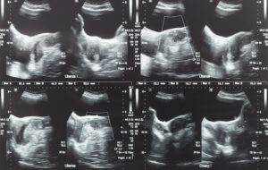
Ultrasound image of lower abdomen, ovary and uterus with tumor or uterine fibroid, leiomyoma of female woman patient for gynecological medical exam, analysis and test

Testing and Diagnosis of Uterine Fibroids

In many cases, especially those whereby uterine fibroids do not present any noticeable symptoms for the patient, this condition will be discovered in a regular pelvic exam. This is a routine checkup carried out by gynecologists. While this checkup might determine the presence of a mass or lump on the uterus, further testing will have to be carried out to make a definite diagnosis.

These testing methods include:

Note: Upon learning that fibroids are a type of tumor, the foremost concern for many patients is whether the tumor is cancerous or might predispose them in some way to developing cancer. Fibroids are rarely cancerous, with the chances of such fibroids (leiomyosarcomas) arising at less than one in a thousand.  Additionally, the presence of fibroids in no way increases a patient’s chances of developing any type of cancer.


What causes uterine fibroids?2022-04-29T17:20:54-07:00

What are Uterine Fibroids?

2022-04-29T17:17:06-07:00

Millions of women around the world are affected by the development of uterine fibroids each year. While it is not a life-threatening condition in itself, the symptoms and potential complications that come with it make it such a pressing medical concern for affected individuals.

uterine fibroids

As only women will usually possess a uterus, it follows that only women are afflicted by this ailment, which is why gynecologists will often be the first ones to make a definite diagnosis. As with so many medical emergencies, knowing the problem early will greatly increase the chances of beating it, which is why the professionals at La Jolla Vein & Vascular are dedicated to administering reliable, accurate, and entirely effective diagnostics, therapies, and treatments to all clients coming through our doors.

Let’s take a closer look at what exactly we will be dealing with should uterine fibroids turn out to be the issue at hand.

What are uterine fibroids? 

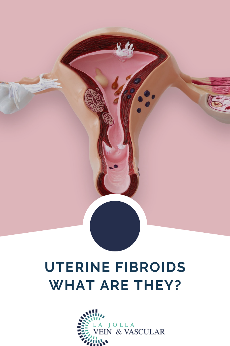
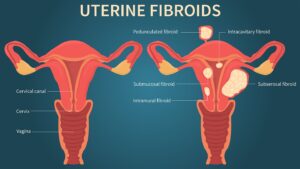
To understand what uterine fibroids are, the first thing one should do is separate the two terms making up the condition. A fibroid is a term to refer to a type of tumor that has ‘muscular’ characteristics. They are also referred to as a leiomyoma and can appear singularly or in formations or in lumps. 

They are known as uterine fibroids since they will appear in the patient’s uterus. Although they are not cancerous in almost all instances, they will range in size from patient to patient, from as small as an apple seed to as large as a grapefruit and beyond, in some unusual instances. 

It is important for all women to be aware of the dangers presented by uterine fibroids, not only owing to the potential harm that the condition might bring about but due to the high rate of prevalence among the female population affected in some way. By the time they are 50 years old, it is estimated between 20 to 80 percent of women will have developed uterine fibroids, with most cases affecting the 40’s to early 50’s. 


Symptoms to look out for: 

Fibroids go undiscovered in most patients due to the lack of symptoms accompanying them, but some patients will experience some symptoms, including:

  • Frequent urination due to pressure being exerted on the patient’s bladder
  • Lower back pain
  • Pain during sexual intercourse
  • Enlargement of the lower abdomen, with severe and extremely rare instances causing the appearance of apparent pregnancy in the patient where none exists
  • Heavy bleeding and/or painful bleeding during periods sometimes resulting in a medically significant depletion of blood in the patient (anemia)
  • Greatly increased risks of complication during pregnancy and delivery, with uterine fibroid patients requiring cesarean sections at six times the average rate.
  • Reproductive health problems such as infertility are very rare outcomes.


For more information on vein and vascular treatment please visit our YouTube Channel.

What are Uterine Fibroids?2022-04-29T17:17:06-07:00

Complications and Causes of Varicoceles

2022-04-29T16:58:30-07:00

What are Varicoceles?

A varicocele is a term used to describe abnormal enlargement of the veins within the scrotum. The veins carry blood from the various body organs back to the heart. Typically, veins have valves that prevent blood backflow. When the valves in the veins within the scrotum don’t work as they should, blood tends to collect in the scrotum, leading to varicoceles.

While varicocele is very common, it is not life-threatening. In fact, research shows that about 15 percent of all adult men have varicocele. For most men, the condition will go unnoticed throughout their life or does not cause any complications. Although the condition is not dangerous, varicocele is a common cause of low sperm production and reduced sperm quality, leading to infertility. Varicoceles can also lead to reduced testosterone production and scrotal discomfort. The condition often does not need treatment until there is a reason for concern about the associated problems. Fortunately, varicocele is easy to diagnose and treat.


Complications and Causes of Varicoceles


Unfortunately, although the condition is not life-threatening, it can also lead to several unpleasant complications.

  • Atrophy (shrinking of the affected testicles)

The testicles are made of sperm-producing tubules. Varicoceles damage these tubules, making the testicles soften and shrink.

  • Infertility

The condition also causes the local temperature around or in the testicles to be too high, affecting sperm formation


Causes

Medical practitioners are not sure of the leading cause of varicoceles. However, doctors agree that the condition forms when there is a problem with blood flow in the spermatic cord. The problem develops when the valves in the veins do not function properly and allows blood to flow back. As a result, blood pools in the veins causing them to dilate. When the condition is common in adults, it can also develop in teens during puberty. The testicles usually require more blood than usual during this growth cycle. This condition can lead to problems in the veins, preventing the blood from flowing where it should.

For more information on vein and vascular treatment please visit our YouTube Channel. 


Complications and Causes of Varicoceles2022-04-29T16:58:30-07:00

La Jolla Vein and Vascular Treatments for Varicoceles

2022-04-29T16:54:19-07:00

La Jolla Vein and Vascular Treatments for Varicoceles 

In most cases, varicoceles treatment is not necessary. Most men with varicoceles do not experience fertility issues. However, if the condition is causing pain, testicular atrophy, or infertility, you can benefit from varicoceles treatment. The treatment helps in sealing off the affected vein and redirecting the blood flow into normal veins. In case of infertility issues, varicoceles treatment helps in improving the quality of the sperm and sperm count. The procedure that we perform at LA Jolla Vein & Vascular on varicocele is percutaneous embolization of varicocele embolization.

The primary goal for varicocele treatment procedures is to help prevent the backflow of blood from the body to the scrotum, helping in cooling off the testes. Surgical procedures will help restrict the blood flow to the veins leading from the testis to the body. However, not all the veins are sealed off. The vassal vein, which is not subject to the same problems as the other varicose veins, is left open to allow blood to leave the testicles after the procedure.

Some of the clear indications that you need varicoceles repair during adolescence include pain, progressive testicular atrophy, and abnormal semen analysis results. While this treatment helps in improving sperm characteristics, it is not clear whether untreated varicoceles can lead to progressive sperm worsening. The common varicoceles treatment options include (La Jolla Vein & Vascular Only Performs Varicocele Embolization):

Percutaneous Embolization (Varicocele Embolization)

Percutaneous embolism is an advanced procedure performed by a radiologist. The specialist usually makes a small cut into a vein in the groin and inserts a tube. The doctor will use X-ray imaging to guide them to the affected veins and insert a coil or a balloon into it through the tube. The procedure helps in blocking the blood flow to the varicocele, shrinking it gradually. This procedure is also done with general anesthesia. 

Catheter-directed Embolization

Catheter-directed embolization is a non-surgical, outpatient treatment performed by an interventional radiologist using imaging to guide catheters or other instruments inside the body. Through mild IV sedation and local anesthesia, patients are relaxed and pain-free during the approximately two-hour procedure. 

For the procedure, an interventional radiologist makes a tiny nick in the skin at the groin using local anesthesia, through which a thin catheter (much like a piece of spaghetti) is passed into the femoral vein directly to the testicular vein. The physician then injects contrast dye to provide direct visualization of the veins so he/she can map out exactly where the problem is and where to embolize or block the vein, which reduces pressure on the varicocele. By embolizing the vein, blood flow is redirected to other healthy pathways. Essentially, the incompetent vein is shut off internally by preventing blood flow, accomplishing what the urologist does without surgery. 

Efficacy of Embolization for Varicoceles 

Embolization is equally effective in improving male infertility and costs about the same as surgical ligation. Pregnancy rates and recurrence rates are comparable to those following surgical varicocelectomy. In one study, sixty percent conceived were treated for infertility. 

In another study, sperm concentration improved in 83 percent of patients undergoing embolization compared to 63 percent of those surgically ligated. Patients who underwent both procedures expressed a strong preference for embolization. 

Varicocelectomy


Your doctor will make a small 1-inch incision into your scrotum and use a microscope or magnifying glass to see the veins better. The doctor might use local anesthesia to numb the area. General anesthesia can also help you sleep through the procedure.


Laparoscopic Surgery


This procedure involves making a much smaller incision and inserting a tube that holds the surgical tools. The surgeon will also use a special camera to see inside you. The procedure is done under general anesthesia to help you sleep through the procedure. The results from the other treatments are similar, only that the incision is small with laparoscopic surgery.


Open Surgery


The open surgery treatment is usually done on an outpatient basis and using a local or general anesthetic. The surgeon will make a small incision below your groin or abdomen to access the affected vein. Your doctor uses advanced surgical tools such as Doppler ultrasound and a surgical microscope to help guide the procedure. The patient can return to a regular daily routine after two days.


Recovery


In most cases, you can go back to your routine after two days of varicose treatment. However, it is advisable to take it easy. Avoid strenuous activities and exercise for about two weeks.

People who have undergone percutaneous embolization tend to recover faster. While you may need a day or two off from work, you can return to your workout program in 7-10 days. If the treatment procedure is to help with fertility, your doctor will perform an additional test in 3-4 months. You will be able to see improved results in 6-12 months. It is important to note that more than 50 percent of men who have undergone the procedure have restored their fertility. Additionally, these surgical procedures are also crucial for teens as they help in slowing testicular growth.


For more information on vein and vascular treatment please visit our YouTube Channel. 

La Jolla Vein and Vascular Treatments for Varicoceles2022-04-29T16:54:19-07:00
Go to Top