
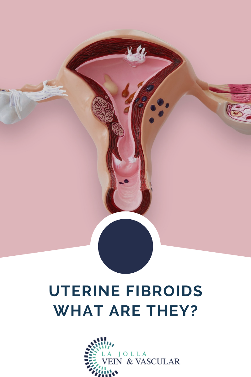
What are Uterine Fibroids?
Millions of women around the world are affected by the development of uterine fibroids each year. While it is not a life-threatening condition in itself, the symptoms and potential complications that come with it make it such a pressing medical concern for affected individuals.


 9")
 10")


