How do you diagnose a DVT? Ultrasound Diagnosis.

Deep vein thrombosis (DVT) poses a significant risk to health, with potentially life-threatening consequences if left untreated. Understanding how blood clots form and identifying the symptoms are crucial steps in managing this condition. But how is DVT diagnosed, and what role does duplex ultrasound play in this process? Let’s delve into the diagnosis of DVT and the significance of duplex ultrasound in its detection and treatment.
Understanding DVT
DVT occurs when a blood clot forms within the deep veins of the leg, thigh, or pelvis, disrupting normal blood flow. While DVT commonly affects the lower extremities, it can occur elsewhere in the body, presenting a range of symptoms and potential complications. Understanding the mechanisms behind blood clot formation and its potential dangers is essential for prompt diagnosis and treatment.
Blood clot formation in the veins typically occurs due to factors such as slow blood flow or pooling, leading to platelets sticking together. While a clot in the deep venous system may not be immediately life-threatening, it can become dangerous if it dislodges and travels to the lungs, causing a pulmonary embolism—an emergency situation that requires immediate medical attention.
Diagnosis of DVT
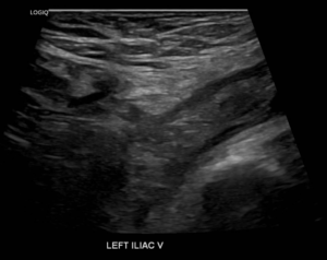
Diagnosing DVT involves a comprehensive assessment by healthcare providers, which may include a physical examination and evaluation of symptoms. The diagnostic approach varies based on the individual’s risk factors and presenting symptoms.
One of the primary tools used in diagnosing DVT is duplex ultrasound, a noninvasive imaging technique that provides detailed visualization of blood flow within the veins. This procedure typically requires no special preparation and is completed within approximately 45 minutes. During the ultrasound, a healthcare provider uses a handheld device called a transducer to capture images of the veins and assess blood flow patterns.
Duplex ultrasound enables healthcare providers to:
- Visualize Clots: By producing real-time images, duplex ultrasound helps identify the presence and location of blood clots within the deep veins of the legs or other affected areas.
- Assess Blood Flow: The ultrasound allows for the evaluation of blood flow dynamics, enabling healthcare providers to detect abnormalities indicative of DVT.
- Monitor Changes: In cases where DVT is suspected but not initially confirmed, repeated ultrasounds over several days may be performed to monitor for the development or progression of blood clots.
Treatment and Management
Upon diagnosis of DVT, prompt intervention is crucial to prevent complications and reduce the risk of further clot formation. Treatment typically focuses on three main goals:
- Preventing Clot Growth: Medications, such as anticoagulants or blood thinners, are commonly prescribed to prevent existing clots from enlarging and to inhibit the formation of new clots.
- Preventing Embolism: By stabilizing existing clots, treatment aims to prevent their dislodgement and migration to the lungs, reducing the risk of pulmonary embolism.
- Preventing Recurrence: Long-term management strategies may involve ongoing anticoagulant therapy and lifestyle modifications to minimize the risk of recurrent DVT episodes.
Duplex ultrasound plays a pivotal role in the diagnosis and management of deep vein thrombosis. By providing accurate and timely assessment of blood flow and clot presence, this imaging technique enables healthcare providers to initiate appropriate treatment strategies, ultimately safeguarding patient health and well-being. Early detection and intervention are key to mitigating the risks associated with DVT, underscoring the importance of regular screening and vigilance in managing vascular health.
“Bringing Experts Together for Unparalleled Vein and Vascular Care”
La Jolla Vein & Vascular (formerly La Jolla Vein Care) is committed to bringing experts together for unparalleled vein and vascular care.
Nisha Bunke, MD, Sarah Lucas, MD, and Amanda Steinberger, MD are specialists who combine their experience and expertise to offer world-class vascular care.
Our accredited center is also a nationally known teaching site and center of excellence.
For more information on treatments and to book a consultation, please give our office a call at 858-550-0330.
For a deeper dive into vein and vascular care, please check out our Youtube Channel at this link, and our website https://ljvascular.com
For more information on varicose veins and eliminating underlying venous insufficiency,
Please follow our social media Instagram Profile for more fun videos and educational information.
For more blogs and educational content, please check out our clinic’s blog posts!

