4, 4, 2022

Smoking and Vascular Disease

2022-04-04T14:53:33-07:00

Smoking and Vascular Disease

The reality of cigarettes is that it is not one product. Every time you smoke, you not only ingest nicotine, but hundreds of other deadly chemicals in the tobacco leaf, paper, contaminants and fertilizers used to grow tobacco. Over time the accumulation of these products causes serious harm to multiple organs. And unlike many things in life, moderation in smoking is not a cure.

Benzene, for example, is a substance on the Environmental Protection Agency’s list of national hazards. It is abundant in tobacco smoke and takes more than 32 hours to completely clear the human body. That means that even ONE cigarette a day continues to keep this poison in your system.

What does smoking do to your arteries?

Smoking is the number one preventable cause of death in the United States. Its effect on heart attacks, peripheral vascular disease and strokes is due to the damage that smoking does to the arteries.

The two chemicals in cigarettes that cause the most trouble are nicotine and carbon monoxide. Nicotine, besides being addictive, has very powerful effects on arteries throughout the body. Nicotine is a stimulant, speeding up the heart by about 20 beats per minute with every cigarette. It raises blood pressure and is a vasoconstrictor, which means it makes arteries all over the body become smaller. That makes it harder for the heart to pump blood through the constricted arteries and it causes the body to release its stores of fat and cholesterol into the blood.

Smoking accelerates the hardening and narrowing process in your arteries; it starts earlier and blood clots are two to four times more likely. Smoking lowers your levels of high-density lipoprotein cholesterol and raises your levels of low-density lipoprotein cholesterol. It decreases the movement of cholesterol through the body, and contributes to its accumulation in your arteries. This puts you at a higher risk for heart attack, stroke, and limb loss.

Your vascular health

Cigarette smoking increases risks of blood clots significantly. If the blood clots in an artery and blood can no longer get through, the tissue that is supposed to be supplied with blood has lost the source of its oxygen and nutrients and dies in minutes. This can result in heart attacks, strokes, and gangrene of the leg.


Smoking effects on the brain:

Smoking increases the risk of stroke by narrowing the arteries in the brain and the carotid arteries in the neck that lead to the brain. In addition, the vessels to the brain can become blocked by a clog or blood clot, which can lead to collapse, stroke and paralysis. If the blood vessels are completely blocked to part of the brain, that part will die. Patients may lose the ability to speak, walk or move normally, or, depending on the part of the brain affected, the stroke may be fatal.


Smoking and Vascular Disease2022-04-04T14:53:33-07:00

29, 3, 2022

What is Peripheral Arterial Disease?

2022-03-29T11:41:41-07:00

What is Peripheral Arterial Disease?

You probably are familiar with the terms angina and heart attack, which are related to a blockage of blood flow to the heart. That same sort of blockage can occur elsewhere in the body, depriving the limbs (the peripheral parts of the body) of oxygen and nutrients. Left untreated, the disease can lead to amputation.

Healthy peripheral arteries are smooth and unobstructed, allowing blood to freely provide the legs with oxygen, glucose and other nutrients. As we age, peripheral arteries build up plaque, a sticky substance made up mostly of fat and cholesterol. Plaque narrows the passageway within the arteries and causes them to become stiff, which then leads into peripheral arterial disease. 

A moderate blockage in one of arteries in a major leg muscle, such as the calf or thigh, can cause pain when walking.This pain can be temporarily decreased with rest, but will act up again with more walking or activity.

Lower extremity pain, similar to angina, is called claudication by medical providers. The pain itself is not limb-threatening but it is a sign that the person should make lifestyle changes and see a doctor. 

Examples of possible treatments for peripheral arterial disease include: 

  • Smoking cessation 
  • Daily exercise
  • Medical management

If the disease becomes more severe, the limb can be starved of basic nutrients and the patient begins to suffer pain at rest (especially at night). Sores that won’t heal may develop on the feet. This is a very concerning sign for losing toes or even amputation of the leg. An urgent referral to a vascular surgeon is necessary to prevent limb loss in this situation.

Causes and risk factors of Peripheral Arterial Disease:

  • Age
  • Gender—males are more prone to the condition than females
  • Hypertension (high blood pressure) 
  • Diabetes
  • Smoking
  • High cholesterol
  • Obesity
  • Lack of exercise
  • Family history of vascular problems

Leg pain does not always indicate PAD, but older patients, especially those who are at risk, should see a physician. A simple, noninvasive ultrasound and leg blood pressure examination can diagnose peripheral arterial disease and determine its severity.

If you experience symptoms of peripheral arterial disease, ask your primary care physician to refer you to a vascular surgeon. Vascular surgeons can perform any necessary treatments, including medication management, minimally invasive endovascular angioplasty and stent procedures, and open bypass surgery.


Treatment for Peripheral Arterial Disease:

Depending on the severity of your condition, treatment options may include lifestyle changes, medications, minimally invasive angioplasty/stenting, or open bypass surgery.


What is Peripheral Arterial Disease?2022-03-29T11:41:41-07:00

Asclera: A New Treatment For Spider Veins and Small Varicose Veins

2022-12-13T13:26:40-08:00

What is polidocanol (Asclera)?

A variety of sclerosant medications can be used for veins, but at La Jolla Vein Care, we prefer polidocanol (brand name Asclera), because it is the most comfortable for the patient and has the lowest risk of side effects. It is a detergent-type medication that injures the inner lining of the veins. This causes the vein to stop flowing, then eventually close down completely. Polidocanol has been well-studied and has been approved by the FDA since 2010 for the treatment of varicose veins and has been used in Europe for decades. Polidocanol is turned into a foam to treat larger veins. This is an off-label use of the sclerosant but very effective and well tolerated. Asclera (polidocanol) Injection is a prescription medicine that is used in a procedure called sclerotherapy to remove unwanted veins on your legs. It is administered by a healthcare provider to treat two types of veins:

Uncomplicated spider veins (very small varicose veins ≤ 1 mm in diameter)
Uncomplicated small varicose veins (1 to 3 mm in diameter) known as reticular veins
Asclera has not been studied in varicose veins more than 3 mm in diameter. La Jolla Vein Care providers have extensive experience with foam sclerotherapy.

Patient Results before and after at 26 weeks after last treatment

asclerabeforeascleraafter

The patient was treated for uncomplicated spider veins (≤ 1 mm)
Treated with 0.5% Asclera

ADVERSE REACTIONS:

-Injection site hematoma
-Injection site irritation
-Injection site discoloration
-Injection site pain
-Injection site itching
-Injection site warmth
-Neovascularization
-Injection site clotting

How often do I need treatment to see results?

The number and frequency of treatments depends on the size of the vessels and their location. Ask your provider about an individualized treatment approach that is right for you.

asclerabeforeafter

How does Asclera work?

Asclera is a sclerosing agent that is injected into the vein. It works by damaging the endothelium, the inside lining of blood vessels. This causes blood platelets and cellular debris to attach to the lining of the vessels. Eventually, cellular debris and platelets cause the blood vessel to clot. Over time, the clotted vein will be replaced with tissue.

How satisfied are patients who use Asclera?

88% of clinical study patients were satisfied or very satisfied with their Asclera treatment after 12 weeks.

ascleragraph
How long is each Asclera session?

A typical session lasts 15 to 45 minutes. Generally, 1 to 3 injections may be necessary to treat a given spider or reticular vein. Repeat treatment sessions may be necessary. Any additional treatment sessions are usually separated by 1 to 2 weeks.

What should I expect after being treated with Asclera?

Following treatment, you’ll need to wear compression stockings day and night for 2 to 3 days, then for 2 to 3 weeks during the daytime, as directed by your provider. Compression stockings are designed to apply pressure to your lower legs, helping to maintain blood flow, and reduce discomfort and swelling. Compression helps your legs heal and is necessary to reduce the risk of deep vein thrombosis.

Are there activities I should avoid after receiving an Asclera Injection?

For 2 to 3 days following the treatment, avoid:
-Heavy exercise
-Sunbathing
-Long plane flights
-Hot baths, hot tubs, or saunas

Possible side effects to Asclera treatments:

Temporary side effects may occur at the site of the injection, including:

-Bruising
-Raised red areas
-Small skin sores
-Darkened skin in the form of lines or spots
-Multiple tiny red blood vessels
-These side effects usually go away within a few days to several weeks. Some side effects may take months or years to resolve.

For more information or to book an appointment for Asclera therapy, please call our office at 858-550-0330.

Asclera: A New Treatment For Spider Veins and Small Varicose Veins2022-12-13T13:26:40-08:00

What is an abdominal aortic aneurysm (AAA)?

2022-03-24T11:15:19-07:00

What is an abdominal aortic aneurysm (AAA)?

The aorta, the largest artery in your body, runs from your heart, down through your chest, and into your abdomen. The abdominal region of the aorta is responsible for delivering blood to your legs, GI tract and kidneys.

An abdominal aortic aneurysm (AAA) occurs when the wall of the aorta progressively weakens and begins to bulge. An AAA may continue to enlarge and eventually rupture if left untreated, causing severe internal bleeding and possibly death. Nearly 200,000 people in the United States are diagnosed with A A A annually; approximately 15,000 die each year from a ruptured AAA.

In addition to concerns about rupture, clots or debris may also develop within an AAA. These substances can be carried to other areas in the body and block circulation, causing severe pain or possibly limb loss if blood flow is cut off for too long.

AAA can be safely treated with early diagnosis; however, most patients have no symptoms.

Causes and Risk Factors:

Most aortic aneurysms are caused by a breakdown in the proteins that provide structural strength to the wall of the aorta. Some risk factors that contribute to this structural breakdown include:

  • Age—individuals over 60 years are most likely to develop the condition
  • Gender—males are more prone to the condition than females
  • History of atherosclerosis (hardening of the arteries) • Family history of AAA
  • Smoking
  • High blood pressure

Symptoms:

Most people feel no symptoms, and an abdominal aortic aneurysm is often detected when tests are conducted for other unrelated reasons. Those who do have symptoms commonly describe back pain, pulsations in the abdomen, groin pain or sometimes sores on the feet.

Diagnosis

If an abdominal aortic aneurysm is suspected, you likely will be referred for an abdominal ultrasound, a painless, safe test that can screen for and measure the size of an AAA. Computed tomographic angiography (CTA) can assess aneurysm size, location and the extent of impact. This study requires exposure to radiation and injection of an intravenous contrast agent. However, a CTA provides valuable anatomic information and can help your vascular surgeon determine the optimal type of repair.

Medicare Screening Benefit

Medicare offers a free, one-time AAA ultrasound screening for qualified seniors as part of their Welcome to Medicare physical during the first 12 months of their enrollment. Men who have smoked sometime during their life and men and women with a family history of AAA are eligible for this benefit.

Treatment

Vascular surgeons can determine the best procedure for you, based on the size of your aneurysm and other factors. For smaller AAAs, you may need to make lifestyle changes, such as quitting smoking and lowering blood pressure. You may be prescribed medication. Your surgeon will have you come back for regular checkups to see if the AAA has changed.

For large AAAs, or those that have been increasing in size over time, repair may be carried out either by placement of an endovascular stent graft or by open bypass grafting.

Be sure to see a board certified vascular surgeon. They have specific training in both minimally invasive and traditional open repairs. The type of repair conducted is very important to long term durability and freedom from rupture.


What is an abdominal aortic aneurysm (AAA)?2022-03-24T11:15:19-07:00

After care for Foam Sclerotherapy

2022-03-21T15:15:18-07:00

After care for foam sclerotherapy

After Treatment Care

You will be instructed on how long to wear compression stockings after treatment, depending on the size of the veins that are treated. Most patients wear thigh-high compression stockings continuously for 1-3 days, then another 7 days.

For the first 24 hours after the procedure, you should not get the area wet. But after, you may shower with the stockings on or take a quick cool shower with them off. Avoid heated showers for one week after the foam sclerotherapy treatment.

You should walk 30 minutes twice daily after treatment and move your legs frequently throughout the day with short walks and/or calf exercises. This avoids pooling of blood in the legs. Avoid prolonged sitting during the day, and when you are sitting have your legs be elevated.

Avoid direct sun exposure during the healing process. The two weeks after treatment. Using sun tan lotion after is recommended.

No swimming in public pools, lakes, or oceans for one week to reduce risk of infection.

It is normal to have aching in the treated veins. This responds well to walking, ice packs, and anti-inflammatory medications such as ibuprofen (Advil, Motrin) and naproxen (Aleve).

What to avoid after foam sclerotherapy treatment

For at least two weeks after treatment, you should avoid strenuous exercise (anything more than a brisk walk), heavy lifting, saunas or hot tubs, and leg massages. All of these dilate the superficial veins and interfere with their healing. You should also avoid airline travel for two weeks after treatment.

Possible side effects after foam sclerotherapy treatment

You will likely have some bruising over the injection sites, then the treated veins may become firm and blue. We recommend patients use the product, RECOVA cream, as this helps reduce swelling, redness, and bruising post procedure.

The skin overlying the treated veins may also develop brownish hyperpigmentation as the blood products within those veins are absorbed by the body; in some patients it can take up to a year to fade.

An uncommon complication is a blood clot within a deep vein. The risk is minimized by using ultrasound guidance and performing calf exercises throughout the procedure.


After care for Foam Sclerotherapy2022-03-21T15:15:18-07:00

What is Radiofrequency Treatment for Varicose Veins?

2022-03-17T10:17:50-07:00

At La Jolla Vein Care, we use a special procedure called radiofrequency treatment for varicose veins. This treatment uses a radiofrequency energy to heat up the wall of the vein. This treatment is used for varicose veins. Varicose veins develop when valves in the veins are damaged and this causes the veins to bulge or twist and stand out in the legs. Radiofrequency ablation treatment closes off the blood flow in the vein that causes the varicose veins. 

radiofrequency treatment for varicose veins


Radiofrequency Ablation for Saphenous Vein Reflux 

Why was I offered an endovenous radiofrequency ablation procedure?

Most patients who are offered a radiofrequency ablation procedure is for backwards flow (or reflux) in the saphenous vein(s). The great and small saphenous veins are the two main superficial veins of the leg. They run along the inner leg and the back of the leg, respectively. This minimally invasive procedure can be performed in the office in less than an hour and patients return to their usual level of activity the same day. 


How does the treatment work?


The skin is numbed with lidocaine, then a tiny wire and the Closurefast catheter are inserted into the vein. The catheter delivers radio-frequency energy to the vein wall, causing it to seal shut. The remaining healthy veins continue to bring blood back to the heart. 


What should I expect on the day of treatment?


The procedure is performed with local anesthesia, but many patients elect to use a mild oral sedative (Valium), which is taken after checking in and completing all paperwork. You will change into a gown and leave underwear on. Depending on the vein to be treated, you will lay on your back or on your belly. We do our best to make special accommodations (for example, if you cannot lie flat or cannot bend a knee very well) with body positioning and using pillows. We will do our best to make you comfortable. Then, we will give you the option of watching a movie on Netflix or listen to music. Once you are comfortable, your leg will be prepped with a cleansing solution for the sterile procedure. The doctor will perform an ultrasound to map the vein to be treated. Then, a numbing agent (lidocaine) will be injected into the skin.

In the numb area of the skin, a tiny puncture is made to pass the radiofrequency catheter. Your doctor will then use a  needle to administer a combination of cool saline and local anesthetic around the vein either in the thigh or calf (depending on which vein is treated). This solution numbs the vein and insulates it from the surrounding tissue. After the numbing solution is applied, the vein is painlessly treated with radiofrequency energy. Once your vein has been treated, we will clean your leg and apply a compression stocking which you will wear for 72 hours continuously. You will walk for 30 minutes prior to getting in your car. 


What should I do after treatment? 


You should walk 30 minutes twice daily after treatment and move your legs frequently throughout the day with short walks and/or calf exercises. This will alleviate discomfort and avoid pooling of blood in the legs. Most of the numbing solution around the vein will be absorbed by your body within a few hours, but you may have a small amount of blood-tinged saline leak from your puncture site onto the gauze. In that case, just remove the gauze and roll your stockings back up. 


What should I avoid after treatment?


For at least two weeks after treatment, you should avoid airline travel to minimize pooling of blood in the legs. You should avoid strenuous exercise (anything more than a brisk walk), heavy lifting, saunas or hot tubs, and leg massages. All of these dilate the superficial veins and interfere with their healing. 


What are the possible adverse effects? 


Expect some bruising over the injection sites, which fades over about two weeks. The skin overlying larger varicose veins may also develop some temporary inflammation and/or brownish hyperpigmentation as the blood products within those veins are absorbed by the body. An uncommon complication is a blood clot within a deep vein.


Before and After Varicose Vein Treatment 

4 BA VV NOR21553 BA VV varicose NOR2149

What is Radiofrequency Treatment for Varicose Veins?2022-03-17T10:17:50-07:00

The Best Way to Treat Spider Veins

2022-03-17T10:12:50-07:00

The best way to treat spider veins is through a procedure called Sclerotherapy. At La Jolla Vein Care we have seen great patient transformations with this type of therapy. If you are interested in knowing if you have spider veins, check this article out. 


Sclerotherapy

Why was I recommended to have sclerotherapy injections?

Spider veins and reticular veins of the legs are most commonly treated with sclerotherapy. Sclerotherapy involves an injection of a medication into the vein. This will cause the vein to collapse and gradually fade away. Sclerotherapy has been used to treat spider veins for decades, but newer solutions such as Asclera allow for spider veins to be treated with minimal discomfort and immediate return to activities.

Vein specialists rarely use saline solutions these days, because alternative solutions are less painful and better tolerated. Sclerotherapy is preferred by most vein specialists over laser because spider veins often have underlying feeder veins that can easily be treated with sclerotherapy, but are not addressed by laser. Many people will require more than one treatment session for optimal results. The national average is 2 to 5 treatment sessions. Treatment sessions are often spaced a month apart, but your health care provider will help determine your customized care plan. Wearing compression stockings after treatment will improve results. 


For larger veins, the medication may be turned into a foam, this is referred to as foam sclerotherapy. Foam sclerotherapy is similar to sclerotherapy of spider veins but instead of a liquid solution, a foamed-solution is injected directly into the vein via a small needle. The solution can be seen on ultrasound monitoring which allows it to be directed into nearby varicose veins painlessly. The veins will seal shut, and gradually be broken down by the body. 


What should I expect on my treatment days?

You will sign your consent form then change into shorts provided by the office. We will clean your skin with alcohol. The sclerosant medication will then be injected into your veins with a fine needle. After your treatment, we will help you into your compression stockings, then you will walk for 30 minutes prior to getting into your car. It is normal for your legs to be achy and tender to the touch after treatment. 


What should I do after treatment? 

You will be instructed on how long to wear compression stockings after treatment, depending on the size of the veins that are treated. Most patients wear thigh-high compression stockings continuously for 1-3 days, then another 7 days. You may shower with the stockings on or take a quick cool shower with them off. 


You should walk 30 minutes twice daily after treatment and move your legs frequently throughout the day with short walks and/or calf exercises. This avoids pooling of blood in the legs. Avoid prolonged sitting during the day. 


It is normal to have aching in the treated veins. This responds well to walking, ice packs, and anti-inflammatory medications such as ibuprofen (Advil, Motrin) and naproxen (Aleve). 


What should I avoid after treatment? 

For at least two weeks after treatment, you should avoid strenuous exercise (anything more than a brisk walk), heavy lifting, saunas or hot tubs, and leg massages. All of these dilate the superficial veins and interfere with their healing. You should also avoid airline travel for two weeks after treatment. 


What are the possible side effects?

You will likely have some bruising over the injection sites, then the treated veins may become firm and blue. 


The skin overlying treated veins may also develop brownish hyperpigmentation as the blood products within those veins are absorbed by the body; in some patients it can take up to a year to fade. Some patients temporarily develop some very fine, pink spider veins in areas where veins have been treated (telangiectatic matting). These usually resolve spontaneously over several weeks but occasionally require additional treatment to clear. If you notice the spider veins have not gone away after treatment, this information may be helpful for you. 

Below are the before and after transformations that we have seen in patient cases with spider veins and sclerotherapy.

1 the best way to treat spider veins  4

The Best Way to Treat Spider Veins2022-03-17T10:12:50-07:00

The Best Treatment for Saphenous Vein Reflux

2022-03-17T10:11:22-07:00

The best treatment for Saphenous Vein Reflux is tailored to the individual 


Fortunately, there are a variety of non-surgical treatment options for saphenous vein reflux, the underlying cause for varicose veins and it’s symptoms. Because vein treatment is not, ‘one treatment fits all,’ the treatment plan should be customized to the patient. Non-surgical options include, laser vein ablation, radiofrequency vein ablation, varithena foam, venaseal an mechanical-chemical ablation, also known as clarivein. This article will specifically discuss features about Clarivein which make it a good option for some patients.

Mechanical-Chemical Ablation (Clarivein) for Saphenous Vein Reflux 

The ClariVein procedure for treatment of backwards flow (or reflux) in your saphenous veins. The great and small saphenous veins are the two main superficial veins of the leg. They run along the inner leg and the back of the leg. This minimally invasive procedure can be performed in the office in less than an hour. This offers patients the benefit of being able to return to their usual level of activity the same day. 


How does the treatment work?

The skin is numbed with lidocaine, then the ClariVein catheter is placed into the unhealthy vein. The catheter closes the vein painlessly by delivering two treatments: 


  1. Mechanical treatment with a tiny rotating wire. 
  2. Chemical treatment with polidocanol. 


This technique is highly effective in closing the vein and only requires one skin puncture, similar to placing an iv. 


What should I expect on the day of treatment? 

The procedure is performed with local anesthesia, but many patients elect to use a mild oral sedative (Valium), which is taken after checking in and completing all paperwork. The patient will change into a gown and leave underwear on. Depending on the vein to be treated, the patient will lay on the back or on the belly. We do our best to make special accommodations (for example, if the patient cannot lie flat or cannot bend a knee very well) with body positioning and using pillows. We will do our best to make the patient comfortable. Then, we will give the option of watching a movie on Netflix or listen to music. Once the patient is comfortable, the leg will be prepped with a cleansing solution for the sterile procedure. The doctor will perform an ultrasound to map the vein to be treated. Then, a numbing agent (lidocaine) will be injected into the skin. In the numb area of the skin, a tiny puncture is made to pass the ClariVein catheter. Once the catheter and rotating wire are in place, the doctor will start the treatment. It is common to feel a tickling or vibrating sensation as the doctor treats the vein. Patients usually have minimal to no pain during this procedure. Once the vein has been treated, the patient will wear compression stockings for 72 hours continuously. The benefits of wearing compression stockings during post surgical recovery are mentioned here


What should I do after treatment? 

You should walk 30 minutes twice daily after treatment and move your legs frequently throughout the day with short walks and/or calf exercises. This will alleviate discomfort and avoid pooling of blood in the legs. It is normal to have aching in the treated veins. This responds well to walking, ice packs, and anti-inflammatory medications such as ibuprofen (Advil, Motrin) and naproxen (Aleve). Compression stockings are recommended for after treatment. 


What should I avoid after treatment? 


For at least two weeks after treatment, you should avoid strenuous exercise (anything more than a brisk walk), heavy lifting, saunas or hot tubs, and leg massages. All of these dilate the superficial veins and interfere with their healing. You should also avoid airline travel for two weeks after treatment. 


What are the possible adverse effects? 

Expect some bruising over the puncture site, which fades over about two weeks. The skin overlying larger varicose veins may also develop some temporary inflammation and/or brownish hyperpigmentation as the blood products within those veins are absorbed by the body. An uncommon complication is a blood clot within a deep vein. 

Videos of the treatment, as well as after care instructions can be found on our website or our Youtube Channel.

The Best Treatment for Saphenous Vein Reflux2022-03-17T10:11:22-07:00

28, 2, 2022

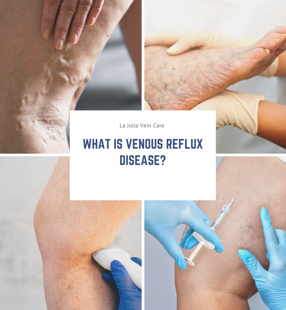
What is Venous Reflux Disease?

2022-03-17T10:21:46-07:00

What is Venous Reflux Disease?


Venous reflux disease is also known as venous stasis, venous insufficiency or venous incompetence and refers to ‘leaky valves’ in the veins of the legs. 
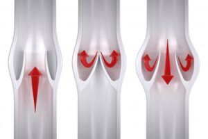
leaky valve

Reflux may occur in the deep and/or superficial leg veins. The deep veins are those within the muscle; they bring at least 80-90% of the blood from the legs back to the heart. The superficial veins are outside the muscle and under the skin. The main superficial veins are the Great Saphenous Vein that courses up the middle of the thigh and calf and small saphenous vein. Normally, there are one-way valves within the leg veins, which help blood flow in one direction: toward the heart. This means blood is traveling against gravity. The calf muscle also helps move blood toward the heart. When vein valves are leaky, blood flows backward (reflux) towards the feet. Blood pools in the lower legs, causing bulging veins at the surface. 

leakyveinvalves

Symptoms of Venous Reflux Disease:


Symptoms of venous reflux disease include; leg heaviness, leg fatigue, leg pain, ankle swelling, phlebitis (inflamed and painful veins), restless legs at night, and night cramps. Venous reflux disease is progressive and worsens over time. Skin changes may also develop, including darkening of the skin around the ankles. The darkening of the skin is sometimes referred to as venous stasis skin changes. The skin can become dry and itchy (venous eczema). Eventually, the skin can break down causing a wound, called a venous leg ulcer. All of which are possible symptoms of venous reflux disease to look out for. 


What influences the development of Venous Reflux Disease?


A patient is more likely to develop venous reflux disease if they are:


  • Overweight
  • Pregnant
  • Have a family history of vein disease
  • Have damage to the leg due to injury, surgery, or previous blood clots
  • High blood pressure
  • Lack of exercise
  • Smoking
  • A blood clot in a deep vein (calf or thigh) “deep vein thrombosis” 
  • Swelling and inflammation of a vein close to the skin, known as “phlebitis”


How is Venous Reflux Disease Diagnosed? 


In addition to physical exam findings and medical history, an ultrasound examination is an important tool in the assessment of venous disease. Not all venous disease is visible to the naked eye, and it usually arises from veins that are beneath the surface of the skin, only visible by ultrasound technology. 


The ultrasound allows us to see if the valves are leaky; it can detect the direction of blood flow and also detects blockages in the veins, for example from blood clots or scars within the veins from previous clots. The ultrasound will determine exactly which veins are “bad” or incompetent. Reflux may be detected in the deep veins (within the muscle), the great and small saphenous veins, and /or branches of the saphenous veins. This will help determine the treatment plan. 


What is Venous Reflux Disease?2022-03-17T10:21:46-07:00

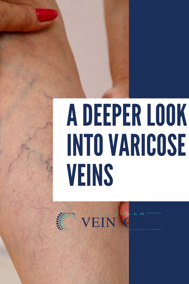
A Deeper Look Into Varicose Veins

2022-03-01T16:31:12-08:00

Varicose veins are swollen, blue, bulging, twisted, superficial (those closest to the skin) veins of the leg. High pressure inside the superficial veins of the leg causes varicose veins, but genetics are thought to play a large, contributing role. As many as 40 million Americans have them. Left untreated, varicose veins may become worse. Persons with varicose veins often experience leg aches and fatigue. They may also incur skin changes such as rashes, redness and ulcers.

What factors influence development of varicose veins?

  • Age: People between the ages of 30 and 70 often have varicose veins. 
  • During pregnancy, 50 to 55 percent of American women experience varicose veins. In most cases, the veins return to normal within a year after childbirth.
  • Women who have multiple pregnancies may develop permanent varicose veins.  


Risk factors include:

o being overweight, or a family history of varicose veins

o standing or sitting for long periods of time


A vascular surgeon will review your medical history, do a physical exam and look at your prominent veins. You may be sent for a duplex ultrasound test, also known as a venous reflux study, to identify the causes of your varicose veins. The painless test uses high-frequency waves to measure the venous blood flow. The test helps the physician visualize the vein structure and the blood flow in the veins.

Most doctors will suggest lifestyle changes for patients. This includes:

  • maintaining the proper weight
  • elevating legs when resting
  • not sitting or standing for long periods of time 
  • wearing compression (elastic support) stockings


In some cases, a vascular surgeon will recommend additional treatments:

  • Sclerotherapy is the sealing of the veins via an injection of a medication. This procedure is done mainly for smaller spider veins. It is performed in a doctor’s office. The physician injects a chemical into the varicose veins to prevent the veins from filling with blood.
  • Vein Stripping is performed. The physician may make two small incisions, one in the groin area and another below the knee, through which the diseased veins are removed. This is an outpatient procedure, usually done under general anesthesia. Patients can resume their normal activities after four weeks.
  • Ablation and laser treatment are performed on patients with severe varicose veins. They are done under local or general anesthetic. The ablation procedure inserts a thin, flexible catheter into the leg vein. The tip of the catheter has tiny electrodes that heat and seal off the walls of the vein. Laser treatments use a tiny fiber placed in the vein through a catheter. The fiber sends out laser energy that closes the diseased portion of the vein. These two modes of treatment frequently replace stripping of the saphenous vein. They can be performed alone or in conjunction with removal of individual clusters of the veins known as small incision avulsion or ambulatory phlebectomy.


A Deeper Look Into Varicose Veins2022-03-01T16:31:12-08:00
Go to Top