25, 7, 2022

Improving your vein health through exercise

2022-12-13T14:12:46-08:00

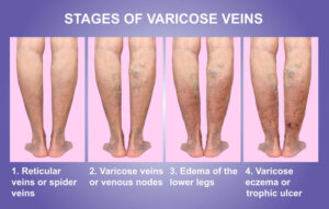
Physical activity and exercise provides a wide range of benefits for vascular health and may help to avoid evolution of mild cases of varicose veins. In fact, exercise is considered to be a fundamental element in improving the symptomatology of patients affected by varicose veins. A sedentary person diagnosed with this medical condition has a much higher risk of worsening the symptoms when compared with an active person with the same condition. This is a result of blood pooling in the veins, causing an increase in venous hypertension and symptoms.

The main goal of exercise in regards to varicose veins is to contract and move the muscles of the leg, helping to pump the blood upwards, avoiding edema or retention of liquid in the ankles. With this in mind, the recommended exercises are those with aerobic characteristics rather than those with anaerobic ones. Through this physical activity, the pressure in the veins is improved, as well as the resulting symptomatology.

Therefore, any exercise that involves moving the lower limbs and promote cyclical muscle contractions is advisable, including stand on tiptoes, move the toes, perform foot bending and rotation, do pedaling movements, among others. These can be easily performed throughout the day without the necessity to go out and exercise, and are especially useful during work hours or while doing daily tasks at home.

When walking or running, pressure is exerted on the sole of the foot, which causes the circulation to be activated from the bottom up, while the constant contraction of the muscles during cycling causes the same effect, but without the presence of high impact, an important factor for those with joint issues.

Swimming is one of the best exercises to practice when affected by varicose veins. The double effect of the water and the movement of the lower limbs cause an incredible increase in blood circulation. This is helpful also for patients who have significant symptoms related to the effects of gravity, like standing.

Other disciplines like yoga, pilates, or rhythmic gymnastics also help stimulate circulation by mobilizing the accumulated blood in the thighs, while relaxing the whole body.

Hiking is a great activity for using the calf-muscle pump. However, in warm weather, symptoms of varicose veins worsen. To get the maximal benefit of exercise and reducing symptoms, outdoor exercising when the weather is cooler, like in the morning is advised.

If you experience any vein disease symptoms, please call our office at (858)-550-0330 to schedule a consultation with one of our knowledgeable doctors at La Jolla Vein and Vascular. 

For more information on vein health please check out our Youtube Channel or visit our helpful guide of resources

Improving your vein health through exercise2022-12-13T14:12:46-08:00

15, 6, 2022

Causes of Osteoarthritis

2022-05-13T17:16:22-07:00

Causes Of Osteoarthritis (OA)


Although Knee Osteoarthritis affects as many as 45 percent of people in their lifetime, the exact cause is unknown. However, medical practitioners have discovered several risks that increase the chances of developing knee osteoarthritis. These include:

Advanced Age

Obesity

Joint Trauma

Family History

Congenital Disabilities or Illnesses

Chronic Injury and Joint Stress

Lack of Exercise

Poor Muscle Tone

Female Sex


Advanced Age

Your susceptibility to developing osteoarthritis increases with age. As you grow old, the cartilage in the knee joint wears down, making it prone to damage and thinning. According to research done by the Arthritis Foundation, about 19 percent of people over 45 years have knee OA. The research further indicates that 37 percent of people over 60 years have the condition, although not all will have the symptoms.


Obesity

The knees support the weight from the thighs upwards. Obese people are at two times more risk of developing knee arthritis than individuals who are not.


Joint Trauma

Surgery, a broken bone, or severe injuries that affect the knee joint can also lead to knee osteoarthritis. These symptoms may not be present immediately after the injuries but may reveal years later.


Family History

Hereditary genes also play a crucial role in the susceptibility of knee arthritis. For instance, if your mother had knee OA, you are more likely to develop the condition than people with no family history of the condition are.


Congenital Disabilities or Illnesses

Septic arthritis, gout, poor bone alignment, congenital conditions, and metabolic disorder can also increase your susceptibility to contracting knee arthritis.


Chronic Injury and Joint Stress

Individuals whose daily activities involve heavy lifting while standing, crawling, or squatting tend to experience mini-traumas. These mini traumas can lead to knee osteoarthritis over time—bodybuilders and other athletes who participate in high-impact sports are prone to knee osteoarthritis development.


Lack of Exercise

Although putting too much stress on your knee joint can lead to arthritis, lack of exercise can also cause OA. Exercise helps in boosting the cartilage’s health and repair. Additionally, the knee joints contain fluid-rich nutrients that need to be circulated in the knee joint capsule. Regular exercise promotes proper circulation of the joint fluid.


Poor Muscle Tone

Poor hamstring, calf, and quadriceps muscle development can put more stress on the knee’s bones and cartilage, causing knee osteoarthritis.


Female Sex

Research shows that women are about 40 percent more likely to develop knee arthritis than men are.


 So, if this is you and you’ve been experiencing pain, please give our office a call at 858-550-0330. 


For more information please check out our Youtube Channel at this link. 


Causes of Osteoarthritis2022-05-13T17:16:22-07:00

30, 5, 2022

What are the risk factors & treatments for uterine fibroids?

2022-04-29T17:24:18-07:00

Who is at Risk of Developing Uterine Fibroids?


There are certain natural circumstances, habits, and lifestyle choices that have been shown to come with an increased chance of developing uterine fibroids, as follow:

Ethnic Origin

Research indicates that women of African American ethnicity are at a significantly higher risk of developing uterine fibroids than Caucasian women for comparison. 

Age

A woman’s advancing age places her at increased risk of developing uterine fibroids. The condition will mostly be found in women in their mid-late forties or early fifties, where the majority enter or pass through their menopausal years. 

Family History

Simple stated, this means that those with a history of uterine fibroids will be more likely to develop the condition. 

Eating Habits

It is believed that the consumption of red meat (beef, ham, etc) in copious amounts will increase the risks of uterine fibroid development. The consumption of green vegetables, on the other hand, is thought to reduce these odds. 

Obesity

Being overweight is considered to be another variable increasing women’s risk of developing this condition, with overweight patients being two to three times more likely to be affected than the average population. 


Treatment Options for Uterine Fibroids

The varied nature of uterine fibroid cases and how they affect different patients in different ways means that dealing with the condition has numerous different approaches. Consultations between patients, whether presenting with symptoms or not, and their physicians should be able to see them agree on a course of action that will satisfy the patient’s best interests. Aside from medication, the typical treatment for fibroids is traditional surgical procedures. At Pedes Orange County, we offer a minimally invasive treatment option that can be reviewed below:

Traditional Surgical Procedures

These are considered invasive surgeries that involve extensive physical intervention. They include:

  • An abdominal myomectomy is an option mostly employed in cases where very large, very deeply embedded, or multiple fibroids are encountered. An open abdominal procedure is often the second-to-last option for patients who see hysterectomies as a last resort.
  • Hysterectomy: This major surgical intervention involves the entire removal of the patient’s uterus and is the only guarantee against the recurrent development of uterine fibroids afterward. While most women will have the option of retaining their ovaries, some will have to undergo hormone replacement therapy if they are not to enter menopause.

Minimally Invasive Surgical Procedure (Uterine Fibroids Embolization by La Jolla Vein & Vascular)

At La Jolla Vein & Vascular, we specialize in Uterine Fibroids Embolization. This minimally invasive surgical procedure involves destroying fibroid masses without necessarily removing them entirely. The techniques are most commonly applied in clinical settings due to their effectiveness, proven track record, and minimal patient impact.

  • Uterine artery embolization: (Also known as Uterine fibroid embolization). Embolic agents (small particles) will be introduced into arteries to block blood flow through them, thus starving fibroids of the nutrients needed for their survival and growth.

In surgical procedures that do not entirely remove the uterus, there will be a remaining risk of new uterine fibroid development in the future.

Why Uterine Artery Embolization (UFE) Instead of a Hysterectomy

Benefits of UFE Over Hysterectomy

Uterine artery embolization (UFE ) is a minimally invasive procedure that effectively treats uterine fibroids and is considered an alternative to uterine fibroid surgery removal. Uterine artery embolization (UFE) is typically performed as an outpatient procedure, which means that most women go home the same day for recovery.

UFE has several benefits over hysterectomy. Besides involving only a small nick in the groin or wrist for catheter insertion, the uterus is preserved. There is no scar with UFE. For a UFE, conscious sedation is used, unlike a hysterectomy, where the woman receives general anesthesia. The recovery time for UFE is generally within 2 weeks, which is significantly faster than the usual 6-week recovery time after hysterectomy.

Women who have a hysterectomy that removed both the uterus and ovaries usually get estrogen replacement therapy (ERT) alone. But women who have only the ovaries removed need both estrogen and progestin because estrogen alone can increase the risk of cancer in the uterus.

The recovery time for UFE is generally within 2 weeks, which is significantly faster than the usual 6-week recovery time after hysterectomy.

The idea of having your entire uterus removed is psychologically daunting for women as some women may still want the option to carry a child, do not want to undergo a general surgery, and/or feel like removing the uterus takes away part of their identity as a female.

Treatment for Uterine Fibroids

With modern medication and care, conditions like fibroid tumors have become more treatable and easier to manage. Uterine fibroid embolization, for instance, is a fairly straightforward procedure used in…


What are the risk factors & treatments for uterine fibroids?2022-04-29T17:24:18-07:00

What are Uterine Fibroids?

2022-04-29T17:17:06-07:00

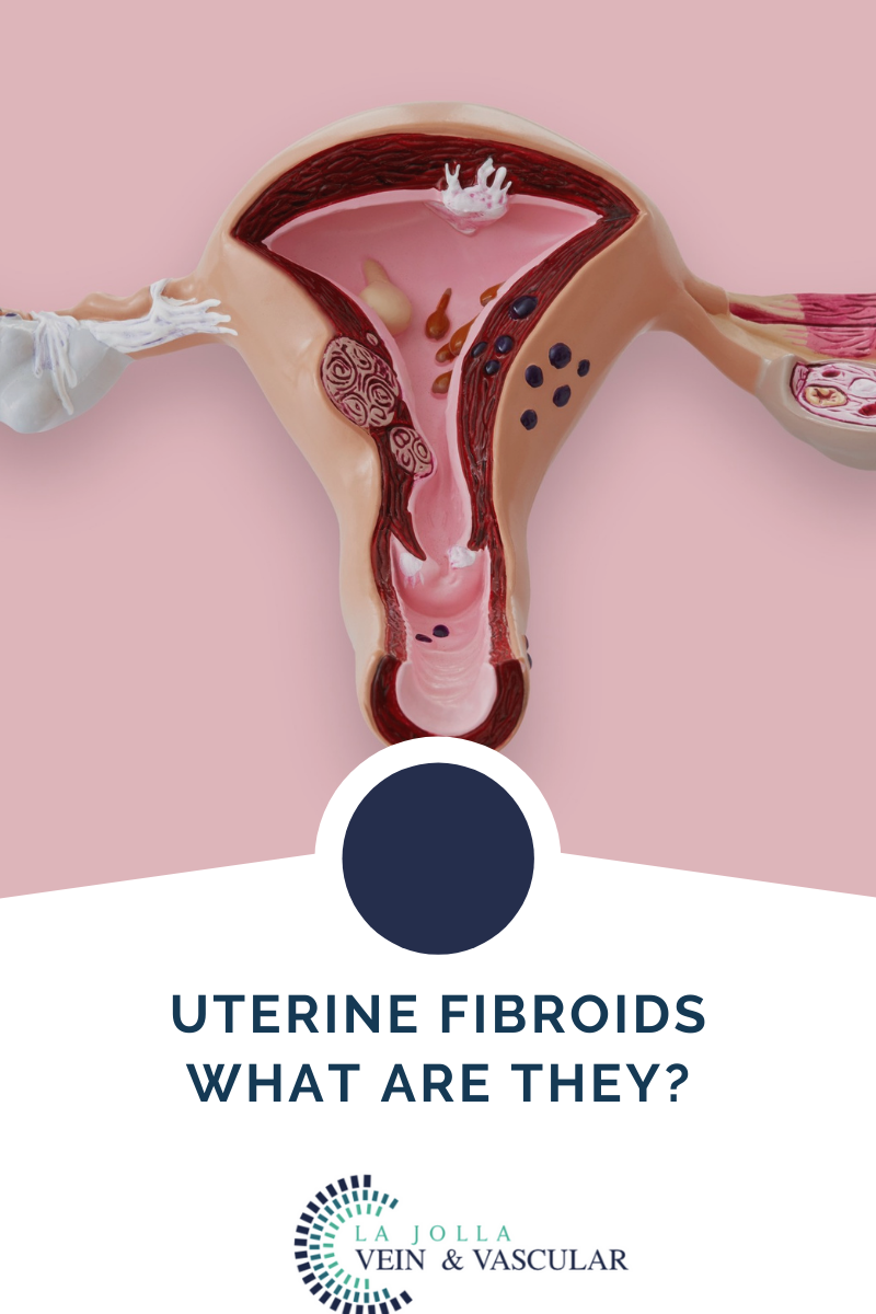
Millions of women around the world are affected by the development of uterine fibroids each year. While it is not a life-threatening condition in itself, the symptoms and potential complications that come with it make it such a pressing medical concern for affected individuals.

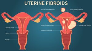
uterine fibroids

As only women will usually possess a uterus, it follows that only women are afflicted by this ailment, which is why gynecologists will often be the first ones to make a definite diagnosis. As with so many medical emergencies, knowing the problem early will greatly increase the chances of beating it, which is why the professionals at La Jolla Vein & Vascular are dedicated to administering reliable, accurate, and entirely effective diagnostics, therapies, and treatments to all clients coming through our doors.

Let’s take a closer look at what exactly we will be dealing with should uterine fibroids turn out to be the issue at hand.

What are uterine fibroids? 

To understand what uterine fibroids are, the first thing one should do is separate the two terms making up the condition. A fibroid is a term to refer to a type of tumor that has ‘muscular’ characteristics. They are also referred to as a leiomyoma and can appear singularly or in formations or in lumps. 

They are known as uterine fibroids since they will appear in the patient’s uterus. Although they are not cancerous in almost all instances, they will range in size from patient to patient, from as small as an apple seed to as large as a grapefruit and beyond, in some unusual instances. 

It is important for all women to be aware of the dangers presented by uterine fibroids, not only owing to the potential harm that the condition might bring about but due to the high rate of prevalence among the female population affected in some way. By the time they are 50 years old, it is estimated between 20 to 80 percent of women will have developed uterine fibroids, with most cases affecting the 40’s to early 50’s. 


Symptoms to look out for: 

Fibroids go undiscovered in most patients due to the lack of symptoms accompanying them, but some patients will experience some symptoms, including:

  • Frequent urination due to pressure being exerted on the patient’s bladder
  • Lower back pain
  • Pain during sexual intercourse
  • Enlargement of the lower abdomen, with severe and extremely rare instances causing the appearance of apparent pregnancy in the patient where none exists
  • Heavy bleeding and/or painful bleeding during periods sometimes resulting in a medically significant depletion of blood in the patient (anemia)
  • Greatly increased risks of complication during pregnancy and delivery, with uterine fibroid patients requiring cesarean sections at six times the average rate.
  • Reproductive health problems such as infertility are very rare outcomes.


For more information on vein and vascular treatment please visit our YouTube Channel.

What are Uterine Fibroids?2022-04-29T17:17:06-07:00

Complications and Causes of Varicoceles

2022-04-29T16:58:30-07:00

What are Varicoceles?

A varicocele is a term used to describe abnormal enlargement of the veins within the scrotum. The veins carry blood from the various body organs back to the heart. Typically, veins have valves that prevent blood backflow. When the valves in the veins within the scrotum don’t work as they should, blood tends to collect in the scrotum, leading to varicoceles.

While varicocele is very common, it is not life-threatening. In fact, research shows that about 15 percent of all adult men have varicocele. For most men, the condition will go unnoticed throughout their life or does not cause any complications. Although the condition is not dangerous, varicocele is a common cause of low sperm production and reduced sperm quality, leading to infertility. Varicoceles can also lead to reduced testosterone production and scrotal discomfort. The condition often does not need treatment until there is a reason for concern about the associated problems. Fortunately, varicocele is easy to diagnose and treat.


Complications and Causes of Varicoceles


Unfortunately, although the condition is not life-threatening, it can also lead to several unpleasant complications.

  • Atrophy (shrinking of the affected testicles)

The testicles are made of sperm-producing tubules. Varicoceles damage these tubules, making the testicles soften and shrink.

  • Infertility

The condition also causes the local temperature around or in the testicles to be too high, affecting sperm formation


Causes

Medical practitioners are not sure of the leading cause of varicoceles. However, doctors agree that the condition forms when there is a problem with blood flow in the spermatic cord. The problem develops when the valves in the veins do not function properly and allows blood to flow back. As a result, blood pools in the veins causing them to dilate. When the condition is common in adults, it can also develop in teens during puberty. The testicles usually require more blood than usual during this growth cycle. This condition can lead to problems in the veins, preventing the blood from flowing where it should.

For more information on vein and vascular treatment please visit our YouTube Channel. 


Complications and Causes of Varicoceles2022-04-29T16:58:30-07:00

Varicocele Diagnosis and Treatments

2022-04-29T16:50:34-07:00

Varicocele Diagnosis


Your doctor will perform a thorough physical examination, which might reveal a non-tender mass above the testicles to diagnose varicoceles. When the mass is large enough, it feels like a bag of worms. When the varicocele is small, the doctor might request you to stand, take a deep breath and hold it while you bear down. This technique will help the doctor to detect any abnormal veins enlargement. Your doctor may also order a scrotal ultrasound. The test uses high-frequency sound waves to create a precise image of the structure of the scrotum. The test is also helpful in helping the doctor rule out other reasons that could be causing the problem in the scrotum.


Treatments for Varicocele

 In most cases, varicoceles treatment is not necessary. Most men with varicoceles do not    experience   fertility issues. However, if the condition is causing pain, testicular atrophy, or infertility, you can benefit.

In most cases, varicoceles treatment is not necessary. Most men with varicoceles do not experience fertility issues. However, if the condition is causing pain, testicular atrophy, or infertility, you can benefit from varicoceles treatment. The treatment helps in sealing off the affected vein and redirecting the blood flow into normal veins. In case of infertility issues, varicoceles treatment helps in improving the quality of the sperm and sperm count. The procedure that we perform at LA Jolla Vein & Vascular on varicocele is percutaneous embolization of varicocele embolization.

The primary goal for treatment procedures is to help prevent the backflow of blood from the body to the scrotum, helping in cooling off the testes. Surgical procedures will help restrict the blood flow to the veins leading from the testis to the body. However, not all the veins are sealed off. The vassal vein, which is not subject to the same problems as the other varicose veins, is left open to allow blood to leave the testicles after the procedure.

Some of the clear indications that you need varicoceles repair during adolescence include pain, progressive testicular atrophy, and abnormal semen analysis results. While this treatment helps in improving sperm characteristics, it is not clear whether untreated varicoceles can lead to progressive sperm worsening. 

The common varicoceles treatment options include (La Jolla Vein & Vascular Only Performs Varicocele Embolization):

Percutaneous Embolization (Varicocele Embolization)

    Varicocelectomy

    Laparoscopic Surgery

    Open Surgery


 Recovery

In most cases, you can go back to your routine after two days of varicose treatment. However, it is advisable to take it easy. Avoid strenuous activities and exercise for about two weeks.People who have undergone percutaneous embolization tend to recover faster. While you may need a day or two off from work, you can return to your workout program in 7-10 days. If the treatment procedure is to help with fertility, your doctor will perform an additional test in 3-4 months. You will be able to see improved results in 6-12 months. It is important to note that more than 50 percent of men who have undergone the procedure have restored their fertility. Additionally, these surgical procedures are also crucial for teens as they help in slowing testicular growth.


Varicocele Diagnosis and Treatments2022-04-29T16:50:34-07:00

Treatment for Non Healing Ulcers and Wounds

2022-04-28T17:49:58-07:00

Treatment of Non-Healing Wounds & Ulcers


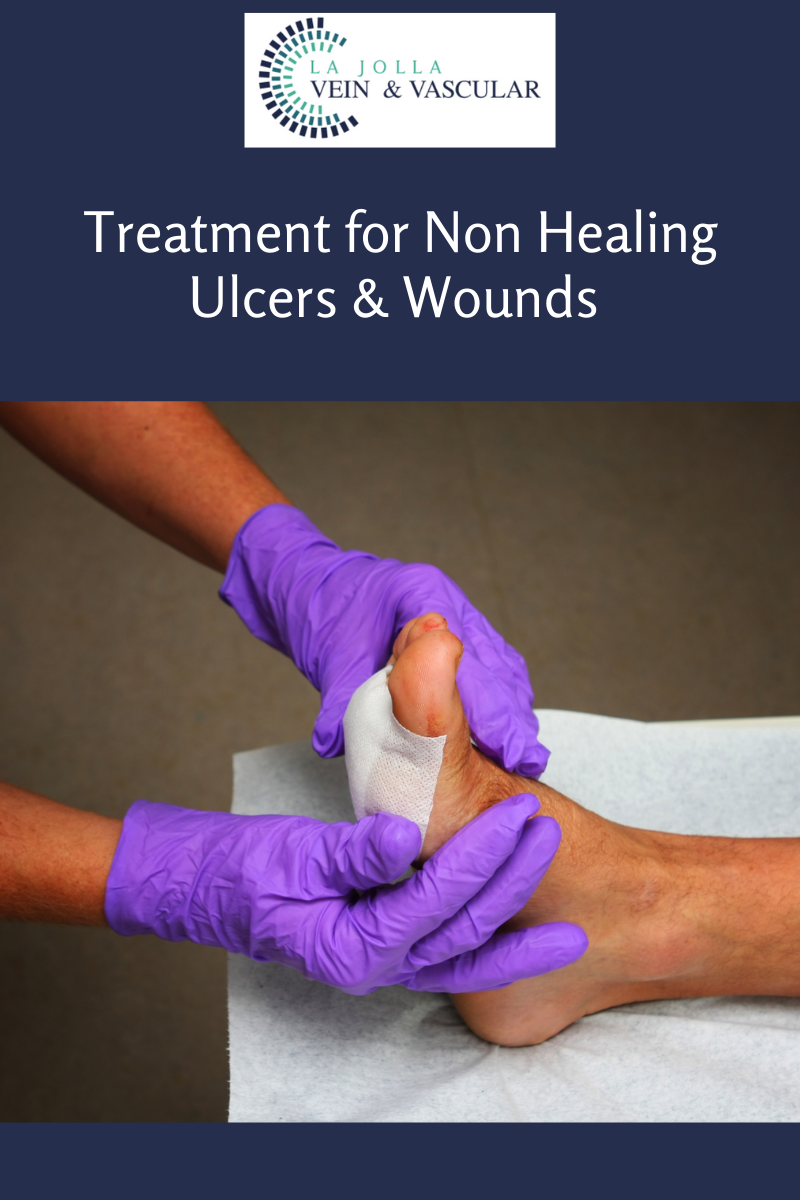
As we’ve seen, there is a very wide range of causes that may contribute to the formation and persistence of non healing ulcers and wounds. It follows that the methods or treatment options at the disposal of healthcare professionals will also be varied. Doctors will discuss the available options with their patients in order to arrive at the best possible options, but the type and severity of the wound will be the decisive point of consideration. These measures include:

Compression Wrapping

Specialized Dressings and Topical Medication

Patient Self-Care and Education

Negative Pressure Therapy (NPWT)

Surgery

Growth Factor Therapy

Debridement (removal of dead tissue)

Compression Wrapping


This involves the application of tightly bound wraps to the affected limbs or regions in order to provide support to the vascular system, which might be operating under lower pressure than it should due to the wearing out of the vascular walls and muscles that support the efficient flow of blood to and fro the tissues that need it.

The doctor will start by doing a thorough physical examination. The doctor will also want to learn about your signs, symptoms, personal health history, risk factors, and family health history. The doctor will ask you several questions regarding your medical conditions, such as diabetes, heart disease, and kidney disease. They will also ask you whether you experience pain or cramps in your leg while walking or exercising.

The doctor will also ask about your family history of PAD and other heart diseases. You will also discuss your smoking habit, either current or in the past. After gathering the information, the doctor will proceed to perform a detailed physical examination. The process involves checking for weak pulses in your leg, listening for poor blood flow in the legs using a stethoscope. The physician will also check for any problems on your legs, such as sores, swelling, and pale skin.

Specialized Dressings and Topical Medication


Technological progress has made it possible to develop special dressing materials made out of smart polymers that work by adjusting their absorptive qualities depending on the hydration levels of the wound area, among other specialized positive actions. Antibiotics may also be employed topically to non-healing ulcers and these help by reducing the bacterial levels present in the wounds in question while maintaining the optimal levels of environmental moisture suitable for wound repair.

ose PAD. This test usually compares the blood pressure in your arm with the blood pressure in your ankle. The doctor usually uses a pressure cuff together with an ultrasound device. Sometimes, the physician may request you to walk on a treadmill and have the doctor take the readings before and immediately after the exercise. The procedure will help the doctor to capture the severity of the narrowed arteries.

Patient Self-Care and Education


There are plenty of measures that patients will be able to implement in the treatment and management if armed with the proper knowledge and aids where required. Regular cleaning and disinfecting of wounds, proper bandage care, manual repositioning of paralyzed limbs, and avoidance of habits that contribute to the incidence or severity of these types of ulcers will be of significant benefit to many.

Negative Pressure Therapy (NPWT)


This treatment method works by pulling away the fluid in the wound that nourishes bacteria by the application of negative pressure. It is also known as vacuum-assisted wound closure for this reason. This action will also reduce tissue swelling and bring up fresh blood and nutrients to the problem area, which assist in the healing processes.


Surgery


In many cases, surgical procedures may be called for to rectify any underlying conditions that might be remedied in this way. Patients with atherosclerosis or blood clots in their vascular system, for example, might undergo surgical procedures to remove these clots or clear built-up cholesterol plaques in their system, thus removing the condition that encourages the formation and persistence of non-healing ulcers.


Growth Factor Therapy


Growth factors refer to the biomaterials generated naturally in our bodies that play the role of replacing tissues as they are damaged or degraded, as in the case of wounds and infections. These factors include epidermal growth factor, insulin-like growth factor (IGF), vascular endothelial growth factor (VEGF), and more. What your medical practitioner will do is introduce or stimulate these factors directly onto the wound in order to speed up the healing and re-growing processes.

Skin Graft Therapy

Skin grafts taken from donors (cadaver, usually) are another option here referred to as allografts. These grafts provide a covering to the wound without integrating with the host body and work by encouraging and providing a structure for the growth of epithelial cells. Severe cases might not be suitable for this intervention, however, and will do better with grafts taken from elsewhere on the patient’s own body.


Debridement (removal of dead tissue)


Debridement more simply refers to the removal of dead or dying (necrotic) tissue from the site. The goal is to minimize the risk or opportunity for infection by eliminating the necessary medium for bacterial growth and proliferation, which such tissue provides. This is an especially relevant measure undertaken in the fight against diabetic non-healing ulcers, as amputation will usually be carried out in cases where infection has progressed past a certain limit.

This is the idea behind the rather outlandish yet scientifically sound application of maggot therapy in the management and treatment of chronic wounds. Here, live, disinfected maggots will be introduced to the wound by a medical professional. Maggots will happily devour necrotic tissue while not touching healthy tissue. This directly leads to a reduction in the bacterial presence in the wound, thus encouraging rapid healing as well as pain and odor mitigation.

La Jolla Vein & Vascular Treatments

The treatment for PAD has two main goals. One is to manage the symptoms, including leg pain, allowing you to resume your normal physical activities. The second one is to top the progression of atherosclerosis in the body


Treatment for Non Healing Ulcers and Wounds2022-04-28T17:49:58-07:00

Symptoms and signs of non healing ulcers and wounds

2022-04-28T17:32:09-07:00

Symptoms of Non-Healing Ulcers


As we’ve seen, there is a very wide range of causes of non healing ulcers and wounds that may contribute to the formation and persistence of non-healing ulcers. It follows that the methods or treatment options at the disposal of healthcare professionals will also be varied.

You shouldn’t allow a wound to fester for weeks on end before seeking professional assistance, especially if you fall under one or more of the risk categories we’ve outlined above. The following are some warning signs to look out for:

  • -Darkening or bluish discoloration around the wound edges
  • -Significant pain around the wound that persists without improvement or progressively grows worse
  • -Foul odor or smell emanating from the wound
  • -Swelling and redness emanating from the wound and spreading to surrounding skin and tissue
  • -Continuous leaking, draining, or weeping from the wound

Notice that these symptoms of non healing ulcers and wounds are mostly indicative of infection. This is a good measure as it often indicates a failure of the body’s natural mechanisms to deal with the wound through its normal processes. The rapid and timely intervention will be called for to forestall further tissue damage and complications.


ulcer4

Treatment of Non-Healing Wounds


Doctors will discuss the available options for non healing ulcers and wounds with their patients in order to arrive at the best possible options, but the type and severity of the wound will be the decisive point of consideration. These measures include:

Compression Wrapping

Specialized Dressings and Topical Medication

Patient Self-Care and Education

Negative Pressure Therapy (NPWT)

Surgery

Growth Factor Therapy

Debridement (removal of dead tissue)

Symptoms and signs of non healing ulcers and wounds2022-04-28T17:32:09-07:00

26, 4, 2022

La Jolla Vein and Vascular welcomes Jodi Hirsch, PA-C

2022-04-23T14:24:17-07:00

La Jolla Vein and Vascular welcomes Jodi Hirsch, PA-C

jodihirsch

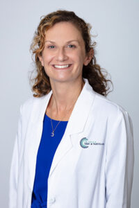
Jodi Hirsch PA-C is a board-certified physician assistant who has spent her career specializing in the field of peripheral vascular disease and venous disease.  She gained her undergraduate degree in chemistry and pursued her degree in Physician Assistant Studies from Touro University in NY in 2006. She has spent her professional career in San Diego, previously working in the Department of Vascular Surgery at Scripps. When not at work, she enjoys spending time with her husband and two little boys.

For more information please watch Jodi’s video here.

La Jolla Vein and Vascular welcomes Jodi Hirsch, PA-C2022-04-23T14:24:17-07:00

Varicose Veins Before & After: Patient Transformations Part 1

2022-04-15T15:34:00-07:00

What are varicose veins?

varicose

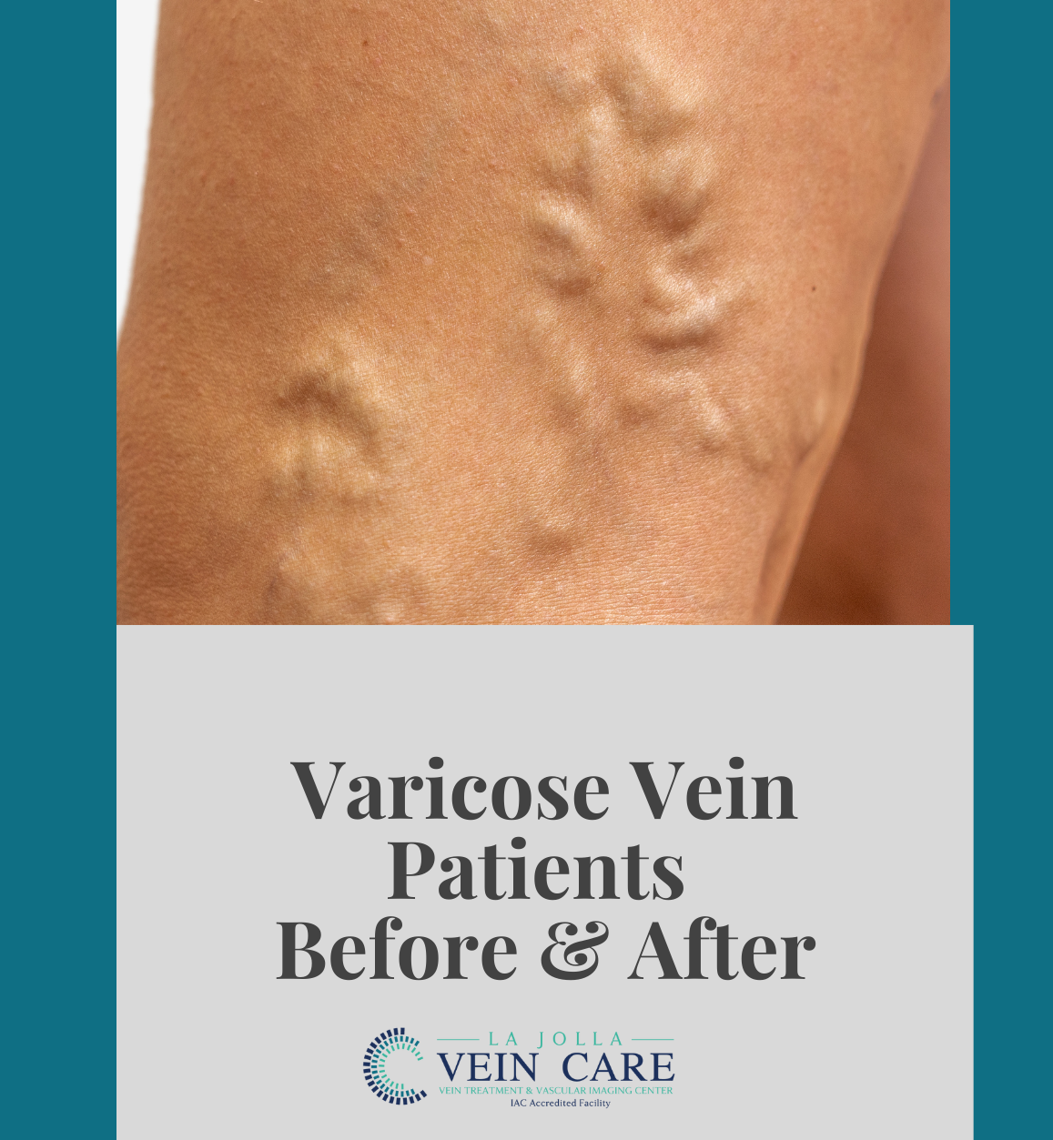
Varicose veins are the twisted, bulging veins just beneath the surface of the skin. Varicose veins are swollen, twisted veins that you can see just under the surface of the skin. These veins usually occur in the legs, but they also can form in other parts of the body.

Varicose veins are caused by leaky vein valves, which allow blood to pool within the veins causing them to stretch and become enlarged. Varicose veins can be an isolated finding, but the majority of the time, they are caused by underlying venous reflux disease. Venous reflux disease is also known as venous stasis, venous insufficiency or venous incompetence. Reflux may occur in the deep and/or superficial leg veins.

In our study, led by Dr. Nisha Bunke and published in the Journal of Vascular Ultrasound in 2018, we studied over 1,000 legs with this vein disease. Over 90% of the time, the source of the varicose veins were the great and small saphenous veins. The Great Saphenous Vein (GSV) courses up the middle of the thigh and calf and the small saphenous vein (SSV), which courses along the back of the calf. Normally, there are one-way valves within the leg veins, which help blood flow in one direction: toward the heart. This means blood is traveling against gravity. The calf muscle also helps move blood toward the heart. When vein valves are leaky, blood flows backward (reflux) towards the feet. Blood pools in the lower legs, causing bulging veins at the surface. 

La Jolla Vein Care Before and After Transformations:


At La Jolla Vein Care, we are here for our patients from beginning to the end. We love seeing the transformations that take place. Below are a few cases from our patient transformations from before varicose vein treatment to after treatment.

pt1

pt2

pt3


Varicose Veins Before & After: Patient Transformations Part 12022-04-15T15:34:00-07:00
Go to Top