30, 3, 2022

What is Carotid Artery Disease?

2022-03-30T16:49:46-07:00

What is carotid artery disease? 

Your carotid arteries are two main arteries that carry blood from your heart, up through your neck, to your brain. Healthy carotid arteries are smooth and unobstructed, allowing blood to flow freely to the brain and provide oxygen, glucose and other nutrients that your brain cells need. Carotid artery disease is a vascular disease.

Typically with age, the carotid arteries build up plaque, made up mostly of fat and cholesterol. Plaque narrows the insides of the arteries and makes them stiff. This process is commonly referred to as “hardening of the arteries,” or atherosclerosis. Carotid artery disease results when the carotid arteries become narrow or obstructed and provide a risk of the plaque traveling to the brain and causing a stroke.

What is a stroke?

Strokes are the third leading cause of death in the United States and the leading cause of perma- nent disability in older Healthy Plaque Build Up adults. Approximately 25% of strokes occur when bits of carotid plaque or clot break off and flow to the brain. If left untreated, carotid artery disease may lead to stroke, where lack of oxygen and other essential nutrients cause damage to the brain. Depending on its severity, a stroke can be fatal.

Symptoms of carotid artery disease: 

  • There may be no symptoms in the early stages of carotid artery disease, and stroke could be the first sign of the condition.
  • Stroke, however, typically has warning signs, referred to as mini-strokes or transient ischemic attacks (TIAs).
  • Mini-stroke symptoms are usually temporary, lasting a few minutes to a few hours, and should be treated as serious medical emergencies requiring immediate treatment because they are strong predictors of future stroke.

Some symptoms of stroke or TIA may include:

– Weakness, numbness, or tingling on one side of the body

– Inability to control movement of a body part

– Loss of vision or blurred vision in one or both eyes

– Inability to speak clearly

– Difficulty talking or comprehending what others are saying

Causes and risk factors of carotid disease:

  • High blood pressure and age
  • Diabetes
  • Smoking
  • High cholesterol and obesity
  • Lack of exercise
  • Family history of hardening of the arteries and/or stroke

Diagnosis of carotid artery disease: 

If your doctor suspects carotid artery disease, it can be confirmed by undergoing a noninvasive duplex ultrasound examination. The ultrasound can also show how severe the disease is. If you experience symptoms of a mini-stroke, seek medical attention immediately. Your physician may then ask you to see a vascular specialist since they are highly trained in every type of vascular procedure.


Treatment:

Depending on the severity of your condition, treatment options may include lifestyle changes, medications, or procedures such as carotid endarterectomy (cleaning out the plaque with surgery), or in select cases, carotid artery stenting. Some patients benefit from surgery even if they are having no symptoms based upon life expectancy and degree of narrowing.


What is Carotid Artery Disease?2022-03-30T16:49:46-07:00

What to expect when you receive Foam Sclerotherapy

2022-03-25T12:49:38-07:00

Ultrasound-Guided Foam Sclerotherapy


Ultrasound-guided foam sclerotherapy works well for treating surface varicose veins and veins that are not visible to the naked eye. The foam medication can be seen on ultrasound, which allows us to administer it precisely and direct it towards nearby varicose veins. Foam sclerotherapy can be safely used for veins that travel close to the skin or nerves. It also travels easily through veins with many twists and turns. 


Foam treatment of varicose veins is not as new as people think. It was originally described as early as 1944.  Foam sclerotherapy is a method for treating varicose veins. It involves injecting a foamed sclerosant medication into unhealthy varicose veins, causing them to eventually dissolve.

image of foam sclerosant

Colleagues in Spain attracted attention of some vein specialists and interest in the use of foam technology in treating venous insufficiency was reawakened. Administration of foamed sclerosant was reintroduced in the early 1990s by Cabrerra, who summarized a broad experience in 1997. By the 1990’s, broad use of diagnostic ultrasound imaging made it possible to monitor foam distribution with ultrasound scanning. Some 40 years earlier, and before the development of ultrasound scanning, foam had been used in Germany to treat varicose veins.  At that time, foam was made by special syringes and its distribution was assessed by touch, instead of ultrasound scanning.

foam

How many treatments will I need?


The number and frequency of treatments depends on a patient’s anatomy, how well the veins respond to each treatment, and the patient’s treatment goals. The national average is 2 to 5 treatment sessions to achieve 80% improvement. Some larger or resistant veins require two or more treatments to respond completely. 


What should I expect on my treatment days?

You will sign your consent form then change into shorts provided by the office. We will clean your skin with alcohol, then use ultrasound to localize the veins. The foam medication will then be injected into your veins with a fine needle. You will elevate your legs on a wedge pillow for approximately 15 minutes. After your foam sclerotherapy treatment, we will help you into your compression stockings, then you will walk for 30 minutes prior to getting in your car. This is normal for your legs to be achy and tender to the touch after treatment. 

For more information on foam sclerotherapy, read this article.


What to expect when you receive Foam Sclerotherapy2022-03-25T12:49:38-07:00

What is an abdominal aortic aneurysm (AAA)?

2022-03-24T11:15:19-07:00

What is an abdominal aortic aneurysm (AAA)?

The aorta, the largest artery in your body, runs from your heart, down through your chest, and into your abdomen. The abdominal region of the aorta is responsible for delivering blood to your legs, GI tract and kidneys.

An abdominal aortic aneurysm (AAA) occurs when the wall of the aorta progressively weakens and begins to bulge. An AAA may continue to enlarge and eventually rupture if left untreated, causing severe internal bleeding and possibly death. Nearly 200,000 people in the United States are diagnosed with A A A annually; approximately 15,000 die each year from a ruptured AAA.

In addition to concerns about rupture, clots or debris may also develop within an AAA. These substances can be carried to other areas in the body and block circulation, causing severe pain or possibly limb loss if blood flow is cut off for too long.

AAA can be safely treated with early diagnosis; however, most patients have no symptoms.

Causes and Risk Factors:

Most aortic aneurysms are caused by a breakdown in the proteins that provide structural strength to the wall of the aorta. Some risk factors that contribute to this structural breakdown include:

  • Age—individuals over 60 years are most likely to develop the condition
  • Gender—males are more prone to the condition than females
  • History of atherosclerosis (hardening of the arteries) • Family history of AAA
  • Smoking
  • High blood pressure

Symptoms:

Most people feel no symptoms, and an abdominal aortic aneurysm is often detected when tests are conducted for other unrelated reasons. Those who do have symptoms commonly describe back pain, pulsations in the abdomen, groin pain or sometimes sores on the feet.

Diagnosis

If an abdominal aortic aneurysm is suspected, you likely will be referred for an abdominal ultrasound, a painless, safe test that can screen for and measure the size of an AAA. Computed tomographic angiography (CTA) can assess aneurysm size, location and the extent of impact. This study requires exposure to radiation and injection of an intravenous contrast agent. However, a CTA provides valuable anatomic information and can help your vascular surgeon determine the optimal type of repair.

Medicare Screening Benefit

Medicare offers a free, one-time AAA ultrasound screening for qualified seniors as part of their Welcome to Medicare physical during the first 12 months of their enrollment. Men who have smoked sometime during their life and men and women with a family history of AAA are eligible for this benefit.

Treatment

Vascular surgeons can determine the best procedure for you, based on the size of your aneurysm and other factors. For smaller AAAs, you may need to make lifestyle changes, such as quitting smoking and lowering blood pressure. You may be prescribed medication. Your surgeon will have you come back for regular checkups to see if the AAA has changed.

For large AAAs, or those that have been increasing in size over time, repair may be carried out either by placement of an endovascular stent graft or by open bypass grafting.

Be sure to see a board certified vascular surgeon. They have specific training in both minimally invasive and traditional open repairs. The type of repair conducted is very important to long term durability and freedom from rupture.


What is an abdominal aortic aneurysm (AAA)?2022-03-24T11:15:19-07:00

EVLA Treatment for Varicose Veins Aftercare 

2022-03-18T15:37:25-07:00

Endovenous laser vein ablation, also known as endovenous laser therapy (EVLA), is a type of treatment for saphenous vein reflux, the underlying cause of most varicose veins. At La Jolla Vein Care, we offer ALL treatment modalities for varicose veins, not just one. Treatment is customized based on a person’s individual pattern of venous reflux (determined by ultrasound examination), personal preference, and personal health history.


What should I do to optimize recovery after treatment? 


Walking is essential to optimize recovery and avoid pooling of blood in the legs. You should walk 30 minutes twice daily during treatment and to do other calf exercises throughout the day. 


Thigh-high compression stockings must be worn continuously for 72 hours after each treatment and for an additional week during the day after the last treatment. This improves your recovery and reduces complications. 


Ice packs can be used as well as anti-inflammatory medications such as ibuprofen (Advil, Motrin) and naproxen (Aleve) for pain relief. 


No strenuous exercise, hot tubs, or leg massages for two weeks to allow veins to heal. 


Avoid airline travel for two weeks to minimize risk of deep vein clots. 


Avoid Saunas as these dilate the superficial veins and interfere with healing.


We look forward to helping you achieve your treatment goals! 

Please reach us at (858) 550-0330 with any questions. 


EVLA Treatment for Varicose Veins Aftercare 2022-03-18T15:37:25-07:00

How is Venous Reflux Disease Treated?

2022-03-17T10:19:57-07:00

How is Venous Reflux Disease Treated? 


Step 1: Identifying the underlying problem

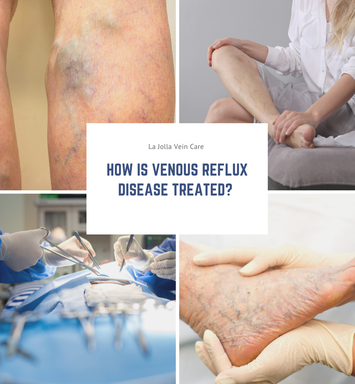
The first step is to treat the underlying problem, venous reflux disease. The specific pattern of venous reflux disease was detected by ultrasound. Venous reflux usually starts in the saphenous veins. The saphenous veins are most effectively treated with vein ablation procedures. This involves placing a small catheter within the vein and using heat or a solution to produce injury and eventful closure of the vein. The most commonly used treatments for the saphenous veins are radiofrequency ablation (RFA), laser ablation, mechanico-chemical ablation (MOCA or Clarivein), and in some cases Varithena Foam. The treatment recommendation is customized, based on where reflux is present and other clinical factors.  

radiofreqgraphic

Step 2: Varicose Veins


After the underlying saphenous vein reflux is corrected, the bulging veins (varicose veins) can be treated by injecting a foamed medication that will cause them to scar and eventually dissolve (foam sclerotherapy), or to remove them using tiny incisions. The most common method is foam sclerotherapy. This is also known as ultrasound-guided foam sclerotherapy (UGFS). Both these treatments can treat venous reflux disease. 


Step 3: Spider Veins 


Spider vein treatment is always considered a cosmetic procedure. If improved cosmesis is a goal of treatment, make sure to discuss this with your doctor and plan on having cosmetic treatment as the last step. Spider vein treatment is accomplished by sclerotherapy, which is an injection of liquid medication into the spider veins. Ask your provider for an estimate about how many treatment sessions/ vials are necessary to get the results you want. 


How is Venous Reflux Disease Treated?2022-03-17T10:19:57-07:00

25, 2, 2022

Can your height influence varicose veins?

2022-02-28T17:01:39-08:00

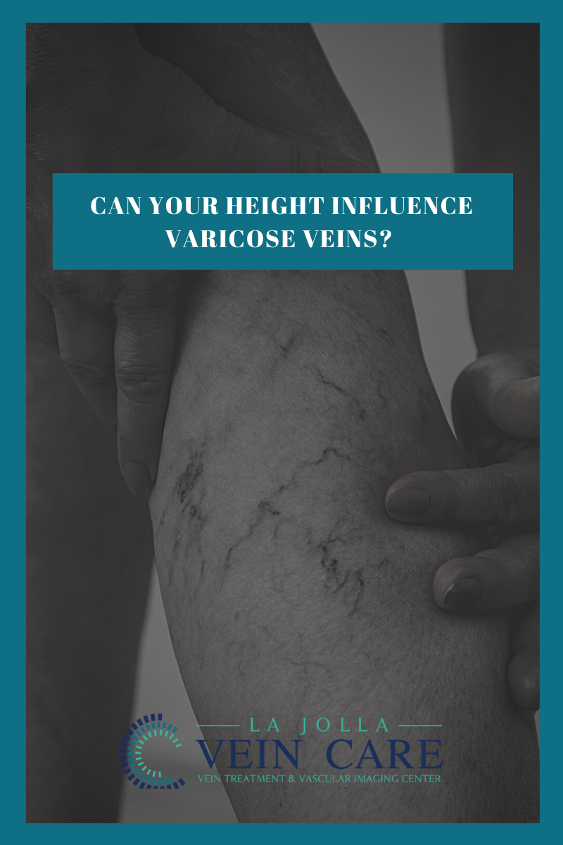
Does height play a role in developing varicose veins? Yes. We initially presented this information in 2018, after a study at Stanford University School of Medicine found a person’s height to be a significant risk factor for developing varicose veins. “We not only found an association between height and varicose veins, but the genetic studies we did showed a causal link,” said cardiologist and study author Dr. Nicholas Leeper, an associate professor of surgery and cardiovascular medicine at Stanford. “That suggests that the genes and pathways that drive human height are also likely to be causing varicose veins.” The study originally published in 2018, found a significant link between above-average height and vein health. In this population-based study of ~500,00 individuals, greater height appeared as a novel predictor of varicose vein disease in machine learning analyses, and was independently associated in multivariable-adjusted Cox regression.

Other known influences such as age, excess weight, being female, and genetics also play a role in the development of them. This study, reveals that taller individuals, the study concluded, have a greater chance of developing them and possibly even blood clots in the legs.

The reason has to do with gravity — blood from the legs of taller people must travel a greater distance to the heart, which forces the veins to work harder. But it also has to do with genetics. The study’s authors found above-average height was not only associated with varicose veins, but could be the underlying cause of the swollen veins that rise to the surface of the legs and feet as well.

Lifestyle modifications to reduce the risk of developing them by including use of compression stockings when traveling and sitting or standing for longer periods of time, exercise, weight loss (if indicated), and leg elevation. Read about measures to reduce symptoms related to varicose veins.

Original Study Published in Circulation.
Clinical and Genetic Determinants of Varicose Veins: A Prospective, Community-Based Study of ~500,000 IndividualsEri Fukaya, Alyssa M. Flores, Daniel Lindholm, Stefan Gustafsson, Daniela Zanetti, Erik Ingelsson, Nicholas J. LeeperCirculation.

Can your height influence varicose veins?2022-02-28T17:01:39-08:00

A Deeper Look Into Varicose Veins

2022-03-01T16:31:12-08:00

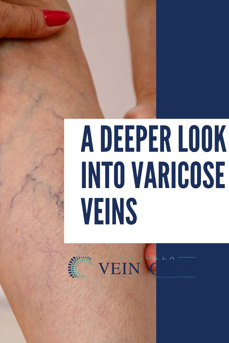
Varicose veins are swollen, blue, bulging, twisted, superficial (those closest to the skin) veins of the leg. High pressure inside the superficial veins of the leg causes varicose veins, but genetics are thought to play a large, contributing role. As many as 40 million Americans have them. Left untreated, varicose veins may become worse. Persons with varicose veins often experience leg aches and fatigue. They may also incur skin changes such as rashes, redness and ulcers.

What factors influence development of varicose veins?

  • Age: People between the ages of 30 and 70 often have varicose veins. 
  • During pregnancy, 50 to 55 percent of American women experience varicose veins. In most cases, the veins return to normal within a year after childbirth.
  • Women who have multiple pregnancies may develop permanent varicose veins.  


Risk factors include:

o being overweight, or a family history of varicose veins

o standing or sitting for long periods of time


A vascular surgeon will review your medical history, do a physical exam and look at your prominent veins. You may be sent for a duplex ultrasound test, also known as a venous reflux study, to identify the causes of your varicose veins. The painless test uses high-frequency waves to measure the venous blood flow. The test helps the physician visualize the vein structure and the blood flow in the veins.

Most doctors will suggest lifestyle changes for patients. This includes:

  • maintaining the proper weight
  • elevating legs when resting
  • not sitting or standing for long periods of time 
  • wearing compression (elastic support) stockings


In some cases, a vascular surgeon will recommend additional treatments:

  • Sclerotherapy is the sealing of the veins via an injection of a medication. This procedure is done mainly for smaller spider veins. It is performed in a doctor’s office. The physician injects a chemical into the varicose veins to prevent the veins from filling with blood.
  • Vein Stripping is performed. The physician may make two small incisions, one in the groin area and another below the knee, through which the diseased veins are removed. This is an outpatient procedure, usually done under general anesthesia. Patients can resume their normal activities after four weeks.
  • Ablation and laser treatment are performed on patients with severe varicose veins. They are done under local or general anesthetic. The ablation procedure inserts a thin, flexible catheter into the leg vein. The tip of the catheter has tiny electrodes that heat and seal off the walls of the vein. Laser treatments use a tiny fiber placed in the vein through a catheter. The fiber sends out laser energy that closes the diseased portion of the vein. These two modes of treatment frequently replace stripping of the saphenous vein. They can be performed alone or in conjunction with removal of individual clusters of the veins known as small incision avulsion or ambulatory phlebectomy.


A Deeper Look Into Varicose Veins2022-03-01T16:31:12-08:00

informacion en español

Visita nuestra página en español

https://ljvascular.com/informacion-en-espanol/

Go to Top