31, 7, 2014

Is Darkening of the Skin A Sign of Venous Disease?

2025-09-04T12:33:20-07:00
cvi3

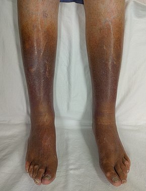
When the skin becomes darker around the ankles, it may indicate underlying venous insufficiency. If you notice that your skin around the ankles changes color, you should see your doctor. Venous insufficiency can be diagnosed with ultrasound scanning by specialists.

Is Darkening of the Skin A Sign of Venous Disease? Yes.   Skin discoloration, also referred to as skin hyperpigmentation or venous stasis, describes the darkening of the skin as a result of venous insufficiency.    This is most frequently cause by venous reflux, an underlying condition that can be treated. When skin changes like these are present, the condition is termed chronic venous insufficiency.  Over time, the skin condition may worsen and the skin may become darker, firm to touch, scaly and itchy, and the skin may break down causing a venous leg ulcer.  It usually occurs around the ankles.  

Is Darkening of the Skin A Sign of Venous Disease?2025-09-04T12:33:20-07:00

Should I Wear Compression When I Travel to Prevent a DVT?

2021-11-05T13:04:03-07:00

Should I Wear Compression When I Travel to Prevent a DVT?

travel

Using compression stockings during travel can reduce the risk of developing a flight-related deep venous thrombosis (DVT).

Yes. Compression stockings are great to use during air travel and long trips where you will be sitting for long periods of time. When you sit for long periods of time you are more at risk of blood clots and swelling. Compression stockings can reduce your risks and prevent swelling.  Ask your doctor the strength that is recommended for you.

Should I Wear Compression When I Travel to Prevent a DVT?2021-11-05T13:04:03-07:00

Why Compression Socks?

2014-07-30T16:21:32-07:00
Meb_Keflezighi

Olympic champion and Boston Marathon winner, Meb Keflezighi wears CEP compression socks while running. CEP compression socks can be found at compressrx.com.

Graduated compression stockings apply external pressure to your legs reducing venous pressure. Graduated compression stockings are great to use if your want to increase circulation, support you leg veins, and want to reduce uncomfortable leg symptoms such as swelling, tired and achy feeling legs.

They are important in the conservative management of varicose veins to reduce symptoms. They are also used after any type of vein treatment to improve the effectiveness of treatment, to reduce bruising and swelling, and for comfort.

Anyone can benefit from compression stockings. Compression stockings are great for relieving ones symptoms when sitting, standing or moving around for long periods of time. They are also great for professionals such as nurses, hairdressers, desk workers, and everyone in between.

Athletes wear compression stockings too.  There is some evidence that medical compression helps to reduce muscle fatigue and improves muscle recovery.  But, not all compression socks are made equal.  They should be medical graduated compression, like CEP socks by Mediven. These are the socks preferred by Olympic champion and Boston Marathon winner, Meb Keflezighi wears CEP compression socks while running.   Find medical CEP compression socks at compressrx.com.


Why Compression Socks?2014-07-30T16:21:32-07:00

Laser and Radiofrequency Vein Treatments

2021-11-04T20:20:33-07:00

What is the difference between laser and radiofrequency procedures for varicose veins?

Both laser and radiofrequency ablation techniques are used as an alternative to surgery for the treatment of varicose veins and underlying venous insufficiency.  The concept behind both laser and radiofrequency treatments is that an energy source is used to heat the vein, causing it to collapse and seal shut. Over time, the treated veins will dissolve. This is offered as an alternative to surgical removal of the veins. The basic difference between the two, are the types of energy sources used.

Laser vein treatment, also known as laser ablation or endovenous laser ablation therapy (EVLT or EVLA) utilizes laser energy to heat the vein.

Radiofrequency vein treatment, also known as radiofrequency ablation (RFA), or the Venefit procedure (previously known as VNUS Closure) utilizes radiofrequency energy to heat the vein.

Both procedures are used to treat the small, great or anterior saphenous veins.

Either procedure involves making a tiny incision the size of a grain of rice on the skin, and a narrow catheter is inserted into the vein. The machine applies either laser or radiofrequency energy to the vein interior, an action that heats the vein and seals it closed.

Unlike traditional surgery, this new minimally invasive procedure takes about 30 minutes to perform and patients can walk out of the office afterward.

While both are effective options for vein treatment, we prefer radio-frequency ablation over laser because it has been proven to be more comfortable than laser yet equally effective. It is also our experience that there is less bruising,  pain, and quicker recovery associated with radio-frequency closure of the veins.

Slide23

Laser vein treatment, also known as laser ablation or endovenous laser ablation therapy (EVLT or EVLA) utilizes laser energy to heat the vein.
Radiofrequency vein treatment, also known as radiofrequency ablation (RFA), or the Venefit procedure (previously known as VNUS Closure) utilizes radiofrequency energy to heat the vein.

Slide24

The radiofrequency catheter is inserted inside the vein. Radiofrequency energy then heats the vein causing it to collapse and seal shut. We prefer radiofrequency over laser vein treatment because we find that it is more comfortable for the patient with less bruising.

Remember, that more than one treatment type is often needed such as radiofrequency of the saphenous vein + sclerotherapy of superficial branches. Talk to your doctor to find out which is best for you or schedule a consultation at 858-550-0330.


Laser and Radiofrequency Vein Treatments2021-11-04T20:20:33-07:00

What is Stasis Dermatitis?

2021-11-05T13:05:00-07:00

Stasis dermatitis or venous stasis dermatitis is a change in the skin that occur when blood collects (pools) in the veins of the lower leg. ‘Stasis’ refers to pooling of the blood in the lower legs from venous insufficiency, and ‘dermatitis’ refers to the inflammation and related skin changes. Because of the inflammation, the skin around the ankles is usually itchy and discolored.

At first, the skin of the ankles and lower legs may look thin or tissue-like. You may slowly get brown stains on the skin.

The skin may become irritated or crack if you scratch it. It may also become red or swollen, crusted, or weepy.

Over time, some skin changes become permanent:

  • Thickening and hardening of the skin on the legs and ankles (lipodermatosclerosis)
  • A bumpy or cobblestone appearance of the skin
  • Dark brown color
Stasis Dermatitis with Ulceration

Chronic venous insufficiency over time can lead to skin changes and eventually ulceration. This image demonstrates stasis dermatitis (also known as venous dermatitis). Stasis dermatitis refers to the skin changes including skin discoloration around the ankles, dry itchy skin that can be thin, and it may eventually break down to cause an ulceration. You need to see a doctor if these signs are present.

Skin sores (ulcers) may develop (called a venous ulcer or stasis ulcer). These most often form on the inside of the ankle.  For this reason, whenever stasis dermatitis or skin changes are noticed around the ankle, you should see your doctor or vein specialist to stop the progression of venous insufficiency and prevent the skin from forming an ulceration.


What is Stasis Dermatitis?2021-11-05T13:05:00-07:00

Varicose Veins vs. Spider Veins

2021-11-05T13:08:51-07:00

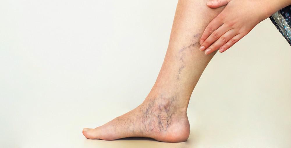
What is the difference between varicose veins and spider veins?  Are they the same thing? Spider veins and varicose veins both refer to dysfunctional, dilated leg veins but the main difference is the size of the veins. Spider veins are small, thread-like veins at the surface of the skin. They often appear in clusters or can have a ‘starburst’ or spider-like pattern. Varicose veins are larger veins that appear swollen, twisted cordlike veins that ‘bulge’ at the surface.

Both spider veins and varicose veins can cause pain and other symptoms like burning, aching, and throbbing. Both can be treated without surgery.

Slide 4_2

This image describes the difference between spider veins and varicose veins. Both are manifestations of unhealthy veins. Spider veins are essential, tiny varicose veins.

Varicose Veins vs. Spider Veins2021-11-05T13:08:51-07:00

How is Venous Reflux Diagnosed?

2021-11-05T11:48:32-07:00


Venous duplex imaging uses ultrasound waves to create pictures.  La Jolla Vein Care utilizes state-of-the-art ultrasound scanners to image the veins beneath the surface of the skin, not visible to the naked eye. Duplex ultrasound imaging can identify if the vein is healthy, or if it is refluxing, or if there are any blood clots in the vein.

Duplex ultrasound combines Doppler flow information and conventional imaging information, sometimes called B-mode, to allow physicians to see the structure of your blood vessels. Duplex ultrasound shows how blood is flowing through your vessels and measures the speed of the flow of blood. It can also be useful to estimate the diameter of a blood vessel as well as the amount of obstruction, if any, in the blood vessel.  Conventional ultrasound uses painless sound waves higher than the human ear can detect that bounce off of blood vessels. A computer converts the sound waves into two-dimensional, black and white moving pictures called B-mode images.

Doppler ultrasound measures how sound waves reflect off of moving objects. A wand bounces short bursts of sound waves off of red blood cells and sends the information to a computer. When performing duplex ultrasound, your ultrasound technologist or physician uses the two forms of ultrasound together. The conventional ultrasound shows the structure of your blood vessels and the Doppler ultrasound shows the movement of your red blood cells through the vessels. Duplex ultrasound produces images that can be color coded to show physicians where your blood flow is severely blocked as well as the speed and direction of blood flow.  Venous reflux refers to back flow of blood across dysfunctional vein valves.  The direction of blood flow is detected by ultrasound.  This is measured in seconds.

US

La Jolla Vein Care image that shows reflux in the great saphenous vein. Duplex ultrasound combines Doppler flow information and conventional imaging information, sometimes called B-mode, to allow physicians to see the structure of your blood vessels.

How is Venous Reflux Diagnosed?2021-11-05T11:48:32-07:00

What is the Femoral Vein?

2022-01-03T13:29:29-08:00

The femoral vein is a large blood vessel of the leg that allows deoxygenated blood to travel to the heart and lungs to become oxygenated. It is located deep within the muscles of the thigh beginning just above the knee (at the adductor canal it is the continuation of the popliteal vein) and ends at the groin level (specifically, it ends at the inferior margin of the inguinal ligament where it becomes the external iliac vein.  It accompanies the femoral artery in the femoral sheath.  In this ultrasound image from La Jolla Vein Care, notice that the femoral vein runs along the same course as the femoral artery, which provides oxygenated blood from the heart and lungs to the rest of the body. The arteries and veins carry blood in opposite directions.  Ultrasound imaging detects the direction of blood flow and in this image, it femoral vein is ‘blue’ depicting blood flow moving toward the heart and the femoral artery is ‘red’ demonstrating blood flow away from the heart.

Ultra Sound detects Femoral Vein

Ultrasound image of the femoral vein and femoral artery. Ultrasound imaging detects the direction of blood flow and in this La Jolla Vein Care image, the femoral vein is ‘blue’ depicting blood flow moving toward the heart and the femoral artery is ‘red’ demonstrating blood flow away from the heart. Notice that the femoral vein and artery are located within the muscle. For orientation purposes, the skin is located at the top of the image.


What is the Femoral Vein?2022-01-03T13:29:29-08:00

8 Warning Signs of Vein Disease: #4 Swollen Ankles

2021-11-05T04:26:19-07:00
After

Ankle swelling related to varicose veins and venous insufficiency may be subtle, leaving indentations from socks.

Swollen Ankles at Night

Thick, swollen ankles are signs that blood or other fluid is congested in the leg and/or leg veins. Over time, damaged vein walls can become even more stretched out and permeable, allowing fluid and protein to filter from the veins into surrounding leg tissue. When you lie down at night, the pressure from gravity is equalized across your leg. Usually, vein related swelling (venous edema) becomes apparent later in the day or worse throughout the day and improved with leg elevation or overnight during sleep.  Often in the morning, there may be no swelling. But, as the day progresses gravity causes poo

Pooling of blood around the ankle in incompetent veins. Venous insufficiency is one of the most common causes of ankle swelling that worsens throughout the day. Sometimes, the swelling can be subtle, leaving indentations from sock lines. Or, it may cause the skin to feel firm, shiny, or puffy by the end of the day.

If you have swelling, you should discuss it with your healthcare provider. Venous insufficiency can be diagnosed with a venous duplex sonogram, which is a non-invasive study of the leg veins.

8 Warning Signs of Vein Disease: #4 Swollen Ankles2021-11-05T04:26:19-07:00

8 Warning Signs of Vein Disease: #3 Open Sores or Ulcers on the Lower Leg

2014-07-02T00:16:13-07:00
Before

Venous leg ulcers are the result of chronic venous insufficiency and venous hypertension, which causes the skin to break down.

When chronic venous insufficiency reaches its most serious point, ulcers may appear on the lower leg. These open ulcers are the result of blood leaking into the leg tissue and damaging the skin. Open sores need to be treated by a doctor immediately.

8 Warning Signs of Vein Disease: #3 Open Sores or Ulcers on the Lower Leg2014-07-02T00:16:13-07:00

informacion en español

Visita nuestra página en español

https://ljvascular.com/informacion-en-espanol/

Go to Top