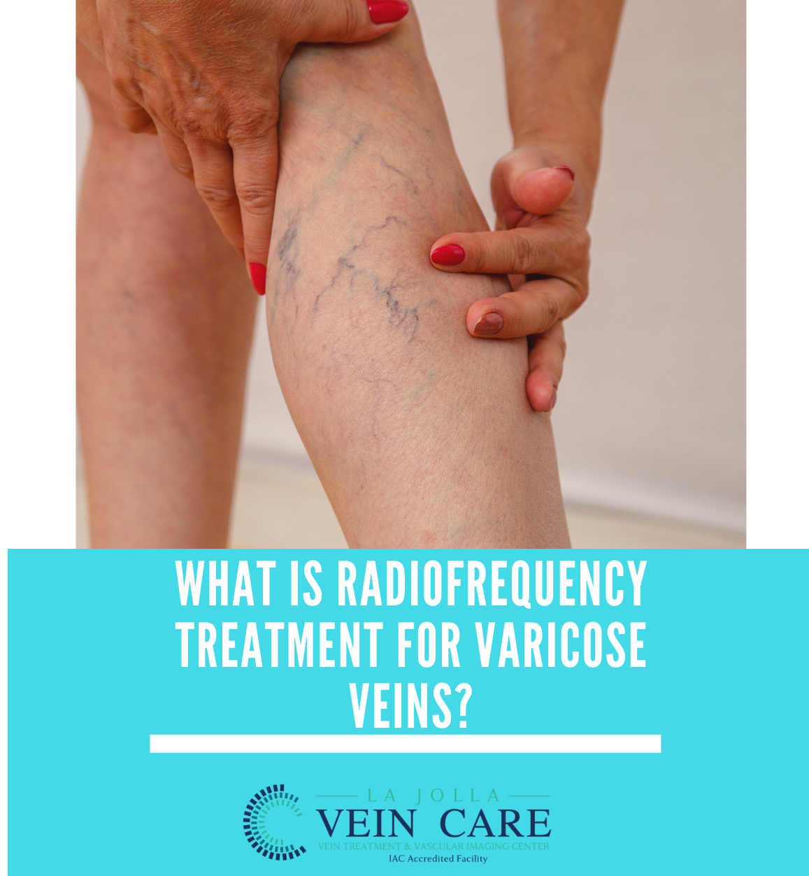
Vascular information from Dr. Bunke

Vein & Vascular News, Tips, Treatments and More!

La Jolla Vein & Vascular welcomes Dr. Elliot DeYoung, M.D.

La Jolla Vein & vascular welcomes our newest doctor at the La Jolla location, Dr. Elliot DeYoung, MD.

new doctor

Dr. Elliot DeYoung is a board-certified interventional radiologist who has spent his career focused on minimally invasive procedures. He completed his training at University of California, San Diego where he specialized in vascular interventions and served on the patient safety committee. Dr. DeYoung’s expertise spans vascular disease from angioplasty and stenting to embolization and ablation. He values thorough patient education and the continuity of patient care.

Dr. DeYoung is excited about joining La Jolla Vein & Vascular team to continue its renowned venous care. He will lead the center in the expansion of vascular services to provide crucial out-patient care to the community. The practice is enthusiastic to offer treatments for peripheral arterial disease, may thurners, and uterine fibroids.

Dr. DeYoung received his bachelor’s degree from USC in Psychology followed by his medical degree from the University of Sydney, School of Medicine. He initially trained as a General Surgery resident in New York before transitioning his training to Interventional Radiology in San Diego. He has a passion for water sports and his free time is spent enjoying the outdoors with his family.

Dr. DeYoung is looking forward to treating your vascular needs at La Jolla Vein & Vascular.

Dr. DeYoung is a vascular specialist with focus on aortic and peripheral vascular disease, pelvic congestion syndrome, and interventional oncology. His skills include varicocele embolization, venous interventions, uterine fibroid embolization, prostate artery embolization, and genicular artery embolization.

Please check out Dr DeYoung’s video for more information.

La Jolla Vein & Vascular welcomes Dr. Elliot DeYoung, M.D.2022-04-23T14:16:23-07:00

A deeper look into chronic venous insufficiency

What is chronic venous insufficiency?

Chronic venous insufficiency (CVI) is an inflammatory condition caused by long standing, untreated venous reflux disease.  Commonly, venous reflux disease causes a collection of symptoms such as leg swelling, heaviness, fatigue and varicose veins. But, over time, when blood is no longer circulating properly in the leg veins, skin changes can also appear. When skin changes appear, this is termed, chronic venous insufficiency (CVI). Venous stasis is another term used for this condition.   Chronic inflammation from CVI causes the skin around the ankles to darken, become dry, itchy, and firm. In severe cases, the skin can start to break down and ulcerate. The ulcer (wound) occurs in the inner or outer ankle and is also referred to as a stasis ulcer or venous ulcer.

Venous stasis skin changes associated with venous insufficiency include:

  • Darkening of the skin along the ankles
  • Itching and dry skin around the ankles (venous eczema)
  • Development of wounds around the ankles, called venous leg ulcers


Chronic Venous Insufficiency is progressive and worsens over time.

Chronic Venous Insufficiency is also known as venous stasis, or venous incompetence. Venous reflux disease refers to ‘leaky valves in the veins of the legs. Reflux may occur in the deep and/or superficial leg veins. The deep veins are those within the muscle; they bring at least 80-90% of the blood from the legs back to the heart. The superficial veins are outside of the muscle and under the skin. The main superficial veins are the Great Saphenous Vein that courses up the middle of the thigh and calf and the small saphenous vein, which courses up the back of the calf. Normally, there are one-way valves within the leg veins, which help blood flow in one direction: toward the heart. This means blood is traveling against gravity. The calf muscle also helps move blood toward the heart. When vein valves are leaky, blood flows backward (reflux) towards the feet. Blood pools in the lower legs, causing bulging veins at the surface. Symptoms include leg heaviness, leg fatigue, leg pain, ankle swelling, phlebitis (inflamed and painful veins) , restless legs at night, and night cramps. Venous reflux disease is progressive and worsens over time. Skin changes may also develop, including darkening of the skin around the ankles. The darkening of the skin is sometimes referred to as skin changes. The skin can become dry and itchy (venous eczema). Eventually, the skin can break down causing a wound, called a venous leg ulcer. 


A deeper look into chronic venous insufficiency2022-04-15T15:36:06-07:00

Understanding Venous Stasis Dermatitis

Understanding Venous Stasis Dermatitis

Venous stasis dermatitis is a change in the skin that occur when blood collects (pools) in the veins of the lower leg. ‘Stasis’ refers to pooling of the blood in the lower legs from venous insufficiency, and ‘dermatitis’ refers to the inflammation and related skin changes. Because of the inflammation, the skin around the ankles is usually itchy and discolored.

In venous stasis dermatitis, the skin of the ankles and lower legs may look thin or tissue-like. You may slowly get brown stains on the skin.The skin may become irritated or crack if you scratch it. It may also become red or swollen, crusted, or weepy.Over time, some skin changes become permanent. Thickening and hardening of the skin on the legs and ankles (lipodermatosclerosis)

Here’s what to look out for:

  • A bumpy or cobblestone appearance of the skin
  • Dark brown color

Venous stasis can cause skin sores (ulcers) may develop (called a venous ulcer or stasis ulcer). These most often form on the inside of the ankle. For this reason, skin changes are noticed around the ankle, you should see your doctor or vein specialist to stop the progression of venous insufficiency and prevent the skin from forming an ulceration.

  Stasis dermatitis

Chronic venous insufficiency over time can lead to skin changes and eventually ulceration. This image demonstrates stasis dermatitis (also known as venous dermatitis). Stasis dermatitis refers to the skin changes including skin discoloration around the ankles, dry itchy skin that can be thin, and it may eventually break down to cause an ulceration. You need to see a doctor if these signs are present.

Eight symptoms to watch out for:

  1. Swelling around ankles
  2. Heavy legs
  3. Varicose Veins, which can be twisted, bulging, dark, or purple
  4. Itching
  5. Pain
  6. Sores that ooze, crust or become scaly
  7. Thickened skin around ankles or your shins
  8. Hair loss on ankles and shins
Understanding Venous Stasis Dermatitis2022-04-15T15:36:58-07:00

Labial Veins and Pelvic Pain May Be Signs of Vascular Disease

Pelvic vein disorder, also known as pelvic congestion syndrome (PCS), is a common disease of the vascular system in women. Pelvic vein disease is a condition in which varicose veins develop in the veins of the pelvis. Like varicose veins in the legs, pelvic vein disease results from impairment of the valves in the veins and engorgement of the blood vessels. It is estimated to occur in approximately 15% of women in the US. Instead of flowing properly to the heart, blood pools in the pelvic veins and causes symptoms such as dull pain, heaviness, and pressure. Symptoms include a dull, aching pain in the pelvic region or lower back, irritable bowel and bladder, pain after sexual intercourse, and vulvar or labial varicose veins. You may also notice varicose veins in the groin or in the upper thigh.
Pelvic Varicose veins
PCS is a highly treatable condition. If you have symptoms of PCS or bulging veins in the vulvar region that affect your quality of life, you should consider being evaluated for PCS. PCS is diagnosed by your medical history, physical examination and special imaging using magnetic resonance venogram to see the blood vessels within the pelvis. Often, we will see large clusters of varicose veins surrounding the ovaries.
Dr. Elliot DeYoung treats PCS. Call us if you would like to discuss concerns about pelvic congestion.

Labial Veins and Pelvic Pain May Be Signs of Vascular Disease2022-04-20T09:58:43-07:00

Chronic venous insufficiency and leg ulcers

Stages of Development of Varicose

Chronic venous insufficiency (CVI) is an inflammatory condition caused by long standing, untreated venous reflux disease that causes leg ulcers.  Commonly, venous reflux disease causes a collection of symptoms such as leg swelling, heaviness, fatigue and varicose veins. But, over time, when blood is no longer circulating properly in the leg veins, skin changes can also appear. When skin changes appear, this is termed, chronic venous insufficiency (CVI). Venous stasis is another term used for this condition.  

Chronic inflammation from CVI causes the skin around the ankles to darken, become dry, itchy, and firm. In severe cases, the skin can start to break down and ulcerate. The leg ulcers or (wounds) occur in the inner or outer ankle and is also referred to as a stasis ulcer or venous ulcer.

Venous stasis skin changes associated with venous insufficiency include:

  • Darkening of the skin along the ankles
  • Itching and dry skin around the ankles (venous eczema)
  • Development of wounds around the ankles, called venous leg ulcers

HOW IS CVI TREATED?

The treatment of CVI cannot reverse the skin changes but it can prevent it from worsening and ulceration. In our experience, treatment of underlying venous reflux reduces the inflammatory changes and the skin may become less itchy and dry, but the permanent skin discoloration does not disappear. Treatment involves correction of the underlying venous reflux, which can help decrease leg ulcers.

HOW ARE LEG ULCERS TREATED?

Venous leg ulcers, depending on the severity, can be healed with a combination of correction of the underlying venous reflux, wound care clinic referral, and compression therapy. A detailed ultrasound will determine what specific treatment is needed depending on where the underlying reflux lies (for example, in the saphenous veins, perforator veins and tributaries). Deep vein reflux is also present in many individuals with ulcers.

leg ulcers

4 BA VLU ulcer DSCF9906 fotor

5 BA VLU ulcer foam results 6 fotor

Chronic venous insufficiency and leg ulcers2022-04-15T15:35:10-07:00

Varicose Veins Before & After: Patient Transformations Part 1

What are varicose veins?

varicose

Varicose veins are the twisted, bulging veins just beneath the surface of the skin. Varicose veins are swollen, twisted veins that you can see just under the surface of the skin. These veins usually occur in the legs, but they also can form in other parts of the body.

Varicose veins are caused by leaky vein valves, which allow blood to pool within the veins causing them to stretch and become enlarged. Varicose veins can be an isolated finding, but the majority of the time, they are caused by underlying venous reflux disease. Venous reflux disease is also known as venous stasis, venous insufficiency or venous incompetence. Reflux may occur in the deep and/or superficial leg veins.

In our study, led by Dr. Nisha Bunke and published in the Journal of Vascular Ultrasound in 2018, we studied over 1,000 legs with this vein disease. Over 90% of the time, the source of the varicose veins were the great and small saphenous veins. The Great Saphenous Vein (GSV) courses up the middle of the thigh and calf and the small saphenous vein (SSV), which courses along the back of the calf. Normally, there are one-way valves within the leg veins, which help blood flow in one direction: toward the heart. This means blood is traveling against gravity. The calf muscle also helps move blood toward the heart. When vein valves are leaky, blood flows backward (reflux) towards the feet. Blood pools in the lower legs, causing bulging veins at the surface. 

La Jolla Vein Care Before and After Transformations:


At La Jolla Vein Care, we are here for our patients from beginning to the end. We love seeing the transformations that take place. Below are a few cases from our patient transformations from before varicose vein treatment to after treatment.

pt1

pt2

pt3


Varicose Veins Before & After: Patient Transformations Part 12022-04-15T15:34:00-07:00

Two Venous Reflux Disease Treatments for visible bulging veins


Two Venous Reflux Disease Treatments for visible bulging veins include: 


Asclera, and Phlebectomy. We at La Jolla Vein Care offer a variety of methods for vein disease. Below is some helpful information about both procedures. 


Ultrasound-guided foam sclerotherapy works well for treating surface varicose veins and veins that are not visible to the naked eye. The foam medication can be seen on ultrasound, which allows us to administer it precisely and direct it towards nearby varicose veins. Foam sclerotherapy can be safely used for bulging veins that travel close to the skin or nerves. It also travels easily through veins with many twists and turns.

Asclera for bulging veins:

A variety of sclerosant medications can be used for veins, but we prefer polidocanol (brand name Asclera®) because it is the most comfortable for the patient and has the lowest risk of side effects. It is a detergent-type medication that injures the inner lining of the veins. This causes the vein to stop flowing, then eventually close down completely. Polidocanol has been well-studied and has been approved by the FDA since 2010 for the treatment of varicose veins and has been used in Europe for decades. Polidocanol is turned into a foam to treat larger veins. This is an off-label use of the sclerosant but very effective and well tolerated. La Jolla Vein Care providers have extensive experience with foam sclerotherapy for the treatment of bulging veins. 

How many treatments will I need?

The number and frequency of treatments depends on a patient’s anatomy, how well the veins respond to each treatment, and the patient’s treatment goals. The national average is 2 to 5 treatment sessions to achieve 80% improvement. Some larger or resistant veins require two or more treatments to respond completely.

Phlebectomy Procedure: 

Phlebectomy is the surgical removal of bulging veins though tiny skin incisions. This minimally invasive procedure is performed in the office with local anesthesia. Phlebectomy is also known as micro phlebectomy (because the incisions are tiny) or ambulatory phlebectomy.

WHAT ARE THE OPTIONS FOR TREATING BULGING  VEINS AT THE SKIN SURFACE?

There are two main techniques to treat varicose veins:

1.) Phlebectomy: surgical removal of the veins

2.) Foam sclerotherapy: injection of a medicated foam to close the veins

Sclerotherapy is the least invasive, but patients with large and/or numerous bulging veins may require several sclerotherapy appointments over two or more months. This is because we can only administer a small amount of foam medication per day and larger veins may require two or more injections to close. Larger veins can also become firm and tender after sclerotherapy and/or deposit brownish blood pigments in the skin as the veins are healing.

WHY WAS I OFFERED PHLEBECTOMY?

Patients with bulging varicose veins may prefer to start with micro-phlebectomy removal of the largest veins, then sclerotherapy treatment for the remaining veins. This reduces the number of appointments and accelerates recovery.

Phlebectomy

HOW DOES THE PROCEDURE WORK?

The skin is numbed with lidocaine, then a tiny puncture is made in the skin (about half the size of a grain of rice). We then infuse additional numbing medication around the veins. A small hook is used to bring the vein to the skin surface, then the vein is removed.


Two Venous Reflux Disease Treatments for visible bulging veins2022-04-15T15:32:43-07:00

What is VenaSeal?

The U.S. Food and Drug Administration (FDA) approved the VenaSeal Closure System to permanently treat varicose veins by sealing the affected superficial veins using an adhesive agent.

vseal

The VenaSeal Closure System is a unique, minimally invasive treatment that uses a safe-for-the-body medical glue to quickly and effectively treat varicose veins (venous reflux disease). Using ultrasound, a doctor will guide a tiny catheter through a small access site in the skin and into the diseased area of the vein. Next, the VenaSeal dispenser delivers a very small amount of medical glue to close the vein. Once the affected vein is closed, blood is immediately re-routed through other healthy veins in the leg.

Unlike other treatments, it does not require anesthesia to be injected into the leg via multiple needle sticks (tumescent anesthesia), and because there are no pre-procedures drugs involved, patients can return to their normal activities right after the treatment. Unlike heat-based procedures, with VenaSeal there is no risk of skin burns or nerve damage. VenaSeal usually does not require any post-treatment pain medication or uncomfortable compression stockings.

procedure

The VenaSeal closure system is the only non-tumescent, non-thermal, non-sclerosant procedure that uses a proprietary medical adhesive delivered endovenously to close the vein. This unique approach eliminates the risk of nerve injury when treating the small saphenous vein, which is a risk sometimes associated with certain thermal-based procedures. Clinical studies have demonstrated that the procedure is safe and effective.

Call La Jolla Vein Care at 858-550-0330 to find out if VenaSeal is a good option for your vein treatment!

Check out our youtube channel for more information on VenaSeal.


What is VenaSeal?2022-04-15T11:21:27-07:00

Understanding The Stages of Venous Disease

The six stages of Venous Disease include:

  1. Spider Veins
  2. Reticular Varicose Veins
  3. Venous Nodes
  4. Chronic Venous Insufficiency
  5. Trophic Ulcers
  6. Varicose Eczema

La Jolla Vein Care gives patients the tailored treatment plan for whichever stage they come in with. A diagnostic test is run called the Duplex Ultrasound. This displays a deeper look into the patient’s veins.

Venous reflux disease is also known as venous stasis, chronic venous insufficiency, or venous incompetence. Venous disease refers to ‘leaky valves in the veins of the legs. Reflux may occur in the deep and/or superficial leg veins. The deep veins are those within the muscle; they bring at least 80-90% of the blood from the legs back to the heart. The superficial veins are outside of the muscle and under the skin. The main superficial veins are the Great Saphenous Vein that courses up the middle of the thigh and calf and the small saphenous vein, which courses up the back of the calf.

Normally, there are one-way valves within the leg veins, which help blood flow in one direction: toward the heart. This means blood is traveling against gravity. The calf muscle also helps move blood toward the heart. When vein valves are leaky, blood flows backward (reflux) towards the feet. Blood pools in the lower legs, causing bulging veins at the surface. Symptoms of venous disease include leg heaviness, leg fatigue, leg pain, ankle swelling, phlebitis (inflamed and painful veins) , restless legs at night, and night cramps. Venous reflux disease is progressive and worsens over time. Skin changes may also develop, including darkening of the skin around the ankles. The darkening of the skin is sometimes referred to as skin changes. The skin can become dry and itchy (venous eczema). 

venous stage


Understanding The Stages of Venous Disease2022-04-14T10:28:50-07:00

Spider Veins Before and After Patient Transformation

What are spider veins?

They are the fine, thread-like reddish veins at the surface of the skin, and are often considered a cosmetic issue, but they can be associated with underlying feeder veins, not visible to the naked eye.  Feeder veins are the ‘blue veins’ also known as ‘reticular veins,’ that give rise to spider veins. They may also be a sign of underlying venous insufficiency. For example, they are usually located in the inner ankles and inner thighs can indicate an underlying problem with the saphenous vein. As a result, an ultrasound examination may be recommended to identify and effectively treat the underlying source.

What are the symptoms?

Vein conditions affect people differently. For some, they can be a painless cosmetic concern, and for others they may cause symptoms. The most common symptoms are burning, throbbing, and localized pain. They can also feel hot and itchy and bleed.

What are the causes?

They are caused by the same condition that causes varicose veins. Leaky vein valves allow blood to pool within the veins causing them to stretch and become enlarged. Spider veins on other areas of the body, such as the face, and chest can be caused by sun damage, hormone changes or liver disease. Hormones, such as with pregnancy, birth control or hormone replacement therapy can weaken the vein wall.

Spider veins and reticular veins of the legs are most commonly treated with sclerotherapy. Sclerotherapy involves an injection of a medication into the vein.  This will cause the vein to collapse and gradually fade away. Sclerotherapy has been used to treat spider veins for decades, but modern solutions such as Asclera, allow for spider veins to be treated with minimal discomfort and immediate return to activities.


Below are La Jolla Vein Care patient transformations before and after vein treatment for spider veins. 

spider veins

spiderveins2

spider veins

Spider Veins Before and After Patient Transformation2022-04-13T11:56:42-07:00
Go to Top