
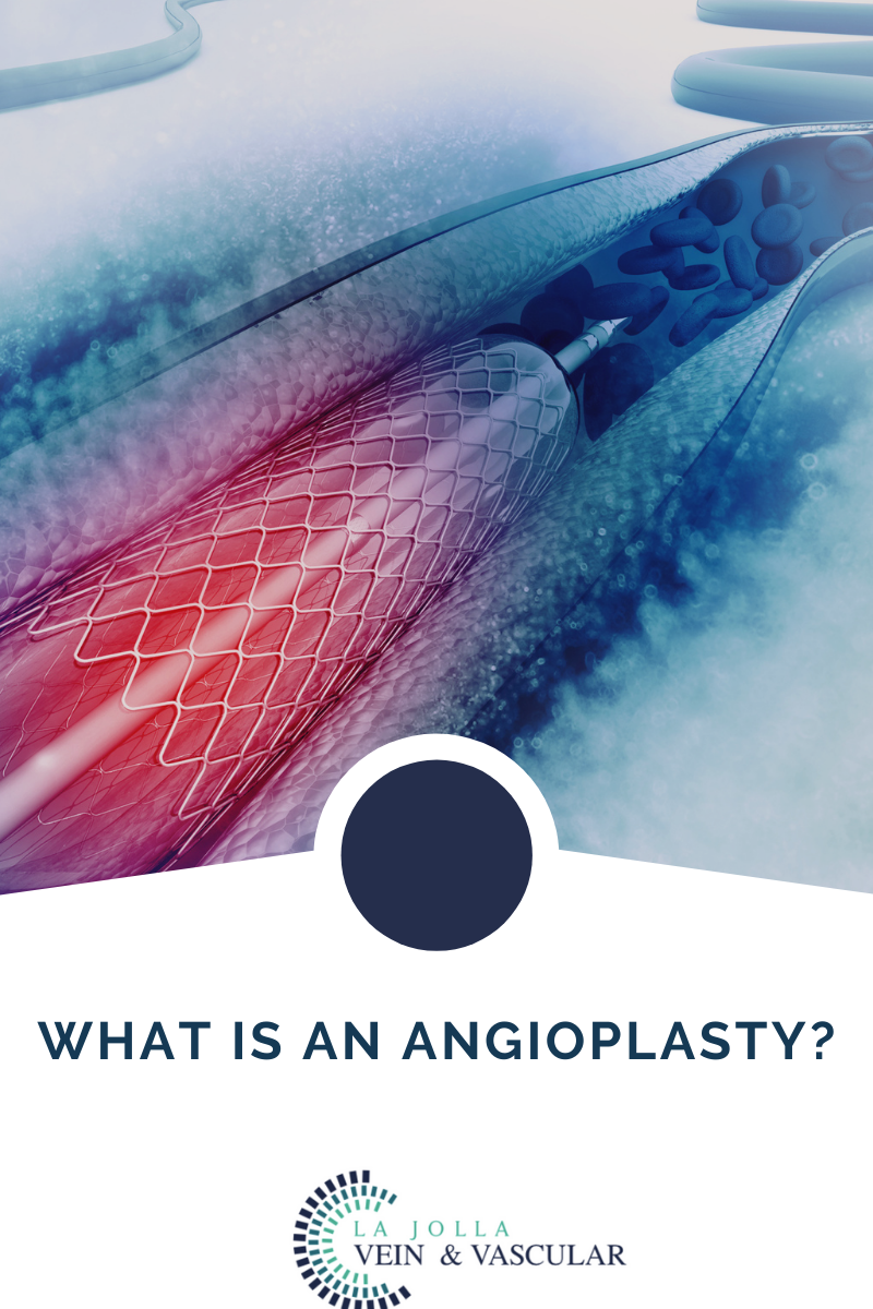
What is an Angioplasty
Angioplasty
At La Jolla Vein & vascular, we are dedicated to offering our patients various procedures and treatment options. One of those options is called an Angioplasty. It is also called percutaneous transluminal Angioplasty (PTA), Angioplasty is a medical procedure performed using a catheter. A catheter is usually a thin, flexible tube inserted through […]


 vs. Hysterectomy 9")
 10")


