What is a Duplex Ultrasound
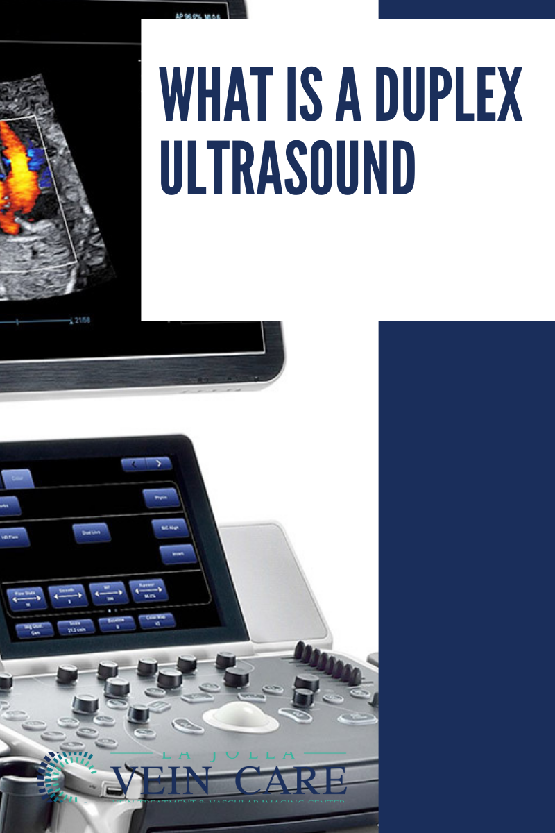
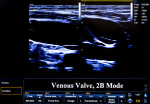
The Duplex Ultrasound examination allows us to visualize the blood vessels that are not visible to the naked eye, even blood vessels that are deep within the muscles. Ultrasound looks at deep and superficial veins in the legs to check for venous-valvular incompetence (the underlying condition that causes varicose veins). The ultrasound examination is used to both identify the veins that have faulty valves and to map the anatomy of the veins, creating a ‘road map.’ This is necessary to make an accurate assessment of the cause and extent of the varicose veins, as well as to formulate the best treatment plan. This should be done for any individual being evaluated for varicose veins, leg swelling, skin changes, patients who have failed prior treatment, patients who are symptomatic and in some patients with certain anatomic patterns of spider veins.
Before your Duplex Ultrasound test:
This study does not require any preparation. You should not wear your compression stockings the same day as the examination. Make sure to be hydrated.
Who Performs the test?
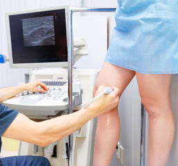
The ultrasound examination is performed by a Registered Vascular Technologist (RVT). An RVT is a sonographer who completed a two-year ultrasound program, plus additional clinical training and obtained certification by meeting the highest standards by The American Registry for Diagnostic Medical Sonography® (ARDMS®). It is important that a specially trained RVT perform the study, because over 40 special images are required to meet accreditation standards. All images are reviewed with the physician.
How long does the duplex ultrasound study take?
Approximately 45 minutes to an hour.
Is it invasive?
No, it is a painless and safe study using sound waves to visualize the veins of the leg.
How to prepare?
This study does not require any preparation. You should not wear your compression stockings the same day as the examination. Make sure to be hydrated.

