How is Venous Reflux Diagnosed?
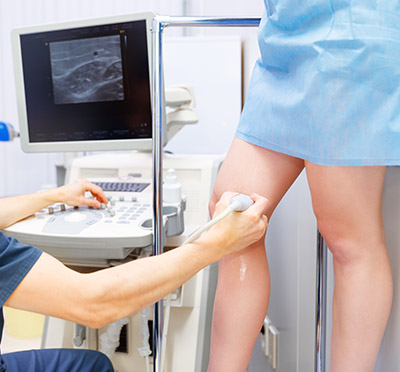
Venous duplex imaging uses ultrasound waves to create pictures. La Jolla Vein Care utilizes state-of-the-art ultrasound scanners to image the veins beneath the surface of the skin, not visible to the naked eye. Duplex ultrasound imaging can identify if the vein is healthy, or if it is refluxing, or if there are any blood clots in the vein.
Duplex ultrasound combines Doppler flow information and conventional imaging information, sometimes called B-mode, to allow physicians to see the structure of your blood vessels. Duplex ultrasound shows how blood is flowing through your vessels and measures the speed of the flow of blood. It can also be useful to estimate the diameter of a blood vessel as well as the amount of obstruction, if any, in the blood vessel. Conventional ultrasound uses painless sound waves higher than the human ear can detect that bounce off of blood vessels. A computer converts the sound waves into two-dimensional, black and white moving pictures called B-mode images.
Doppler ultrasound measures how sound waves reflect off of moving objects. A wand bounces short bursts of sound waves off of red blood cells and sends the information to a computer. When performing duplex ultrasound, your ultrasound technologist or physician uses the two forms of ultrasound together. The conventional ultrasound shows the structure of your blood vessels and the Doppler ultrasound shows the movement of your red blood cells through the vessels. Duplex ultrasound produces images that can be color coded to show physicians where your blood flow is severely blocked as well as the speed and direction of blood flow. Venous reflux refers to back flow of blood across dysfunctional vein valves. The direction of blood flow is detected by ultrasound. This is measured in seconds.

