Ultrasound Findings of Normal vs. Diseased Great Saphenous Vein

2021-11-13T15:00:59-08:00

The Great Saphenous Vein (GSV) is the most commonly affected superficial vein to become diseased (valves no longer function and become leaky).  While venous reflux can involve the deep system and perforators, the superficial venous system is most commonly involved.  The superficial venous system consists of the great saphenous vein (GSV), accessory saphenous veins (AASV), small saphenous vein (SSV), several tributary veins and junctions where they connect to the deep system which include the sapheno-femoral junction (SFJ) and the sapheno-popliteal (SPJ).  Additionally, common anatomical variations of the SSV can occur, such as the presence of the vein of Giacomini (VOG) and a thigh extension (TE).


 GSV imaged

Normal GSV This is the GSV imaged on ultrasound in a longitudinal view. It is healthy without reflux.

GSV with reflux

GSV with reflux

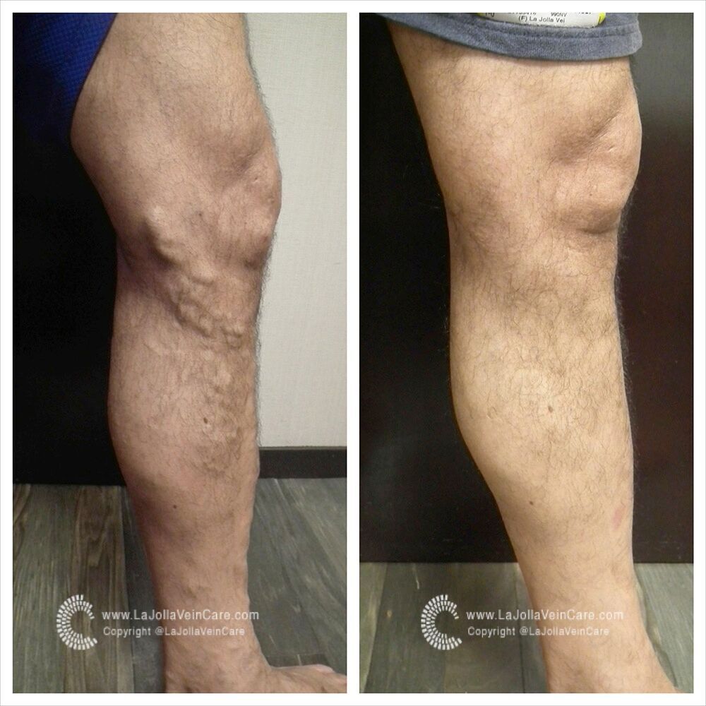
Venous-valvular dysfunction within any of these veins may result in the appearance of varicose veins and can produce a range of symptoms.  Symptoms of venous insufficiency may include leg swelling, aching, heaviness, fatigue that is worse at the end of the day. Common nocturnal symptoms include restless legs and leg cramping. The GSV is most commonly affected, and is most frequently the source of varicose veins. Venous reflux can be determined by ultrasound.