14, 7, 2014

How is Venous Reflux Diagnosed?

2021-11-05T11:48:32-07:00


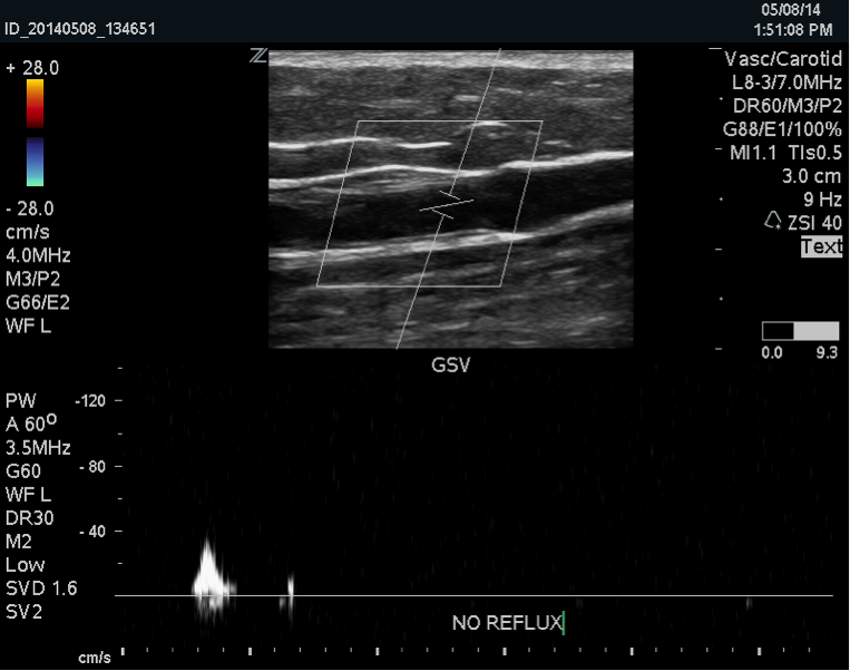
Venous duplex imaging uses ultrasound waves to create pictures.  La Jolla Vein Care utilizes state-of-the-art ultrasound scanners to image the veins beneath the surface of the skin, not visible to the naked eye. Duplex ultrasound imaging can identify if the vein is healthy, or if it is refluxing, or if there are any blood clots in the vein.

Duplex ultrasound combines Doppler flow information and conventional imaging information, sometimes called B-mode, to allow physicians to see the structure of your blood vessels. Duplex ultrasound shows how blood is flowing through your vessels and measures the speed of the flow of blood. It can also be useful to estimate the diameter of a blood vessel as well as the amount of obstruction, if any, in the blood vessel.  Conventional ultrasound uses painless sound waves higher than the human ear can detect that bounce off of blood vessels. A computer converts the sound waves into two-dimensional, black and white moving pictures called B-mode images.

Doppler ultrasound measures how sound waves reflect off of moving objects. A wand bounces short bursts of sound waves off of red blood cells and sends the information to a computer. When performing duplex ultrasound, your ultrasound technologist or physician uses the two forms of ultrasound together. The conventional ultrasound shows the structure of your blood vessels and the Doppler ultrasound shows the movement of your red blood cells through the vessels. Duplex ultrasound produces images that can be color coded to show physicians where your blood flow is severely blocked as well as the speed and direction of blood flow.  Venous reflux refers to back flow of blood across dysfunctional vein valves.  The direction of blood flow is detected by ultrasound.  This is measured in seconds.

US

La Jolla Vein Care image that shows reflux in the great saphenous vein. Duplex ultrasound combines Doppler flow information and conventional imaging information, sometimes called B-mode, to allow physicians to see the structure of your blood vessels.

How is Venous Reflux Diagnosed?2021-11-05T11:48:32-07:00

Foam Treatment of Venous Malformations

2021-11-05T13:22:04-07:00
DSCF0086 copy

Venous malformation of the lateral leg before treatment. Venous malformations appear like abnormally dilated bluish vessels near the surface of the skin. They appear in clusters and look different than typical varicose veins. They often appear during childhood, as opposed to varicose veins.

Venous malformations comprise either superficial or deep veins that are abnormally formed and dilated.  Although they usually are present at birth, they may not be seen until years later into adolescence or even adulthood. The natural history of a venous malformation is slow, steady enlargement. However, events such as surgery, trauma, infection, or hormonal changes associated with puberty, pregnancy or menopause may cause rapid expansion.

At La Jolla Vein Care, we frequently evaluate and treat superficial venous malformations of the legs. Prior to treatment, it is important to have thorough diagnostic imaging, such as an ultrasound examination to ensure an accurate diagnosis of a pure venous malformation.  If an arterial connection (arterial venous malformation) is suspected on ultrasound evaluation, further imaging with CT or MR arteriogram may be necessary. Venous malformations can also occur in other syndromes, such as Klippel -Trenaunay syndrome and may involve the abnormal development of the deep veins.

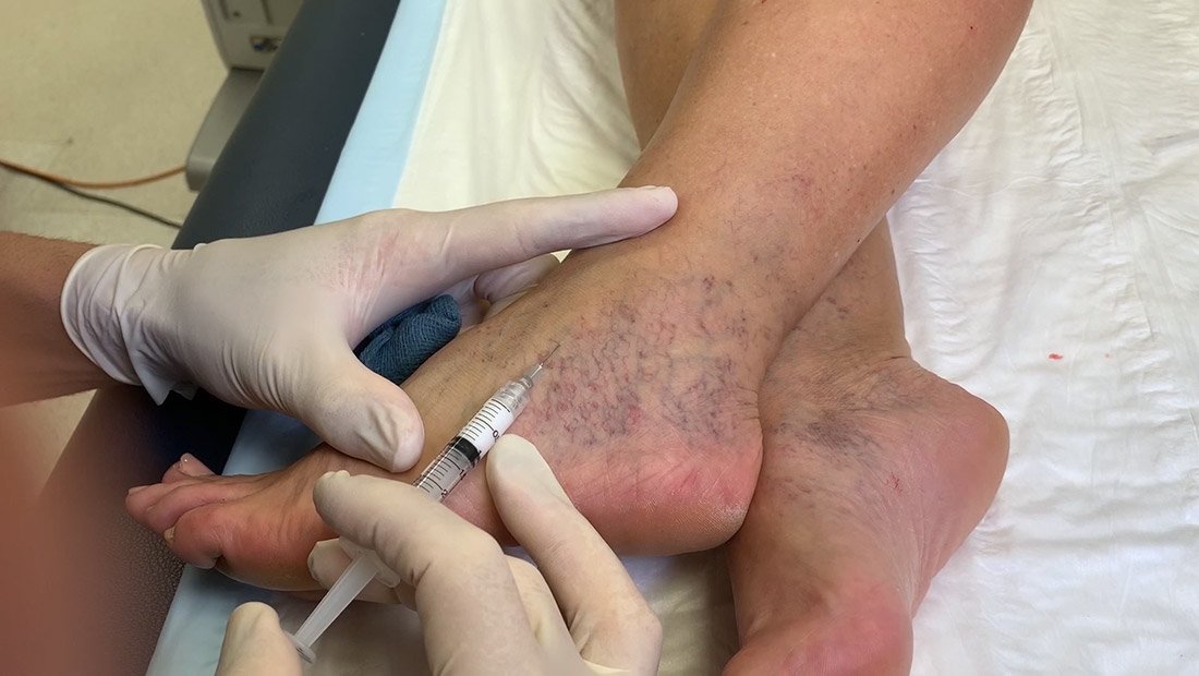
Pure venous malformations can be treated without surgery. Foam sclerotherapy is a common treatment type.  Foam sclerotherapy uses a technique to inject a foamed sclerosing solution into the cluster of varicose veins, which will cause them to collapse and eventually dissolve.

DSCF1899

Venous malformation after treatment with ultrasound-guided foam sclerotherapy at La Jolla Vein Care. Treatment can reduce the symptoms associated with venous malformations such as leg pain, aching and throbbing.

Foam Treatment of Venous Malformations2021-11-05T13:22:04-07:00

18, 6, 2014

History of Foam Sclerotherapy Treatment of Varicose Veins

2021-11-05T10:56:45-07:00
foam

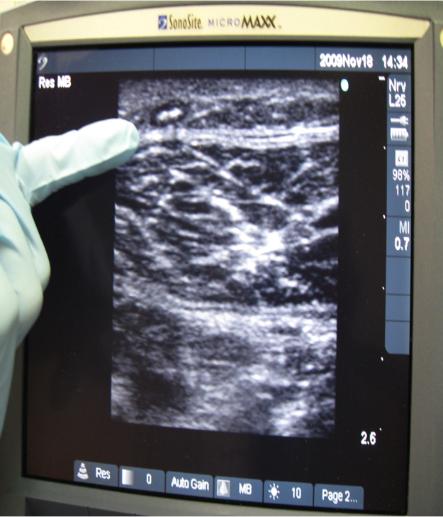
Foamed sclerosant inside a vein: Once foam is introduced into the vein, it is hyperechogenic on ultrasound. In this picture, a La Jolla Vein Care doctor points to the foamed medication inside the vein. Notice it appears ‘white.’

foam03*

Foamed sclerosant used for sclerotherapy has a ‘foam’ or frothy-like appearance.

In 1994 and 1950, E.J. Orbach introduced the concept of a macro bubble air-block technique to enhance the properties of sclerosant in performing macrosclerotherapy.  Apparently, few vascular surgeons were interested in the subject and the technique languished.  The work of Juan Cabrera and colleagues in Spain attracted attention of some vein specialists and interest in the use of foam technology in treating venous insufficiency was reawakened. Administration of foamed sclerosant was reintroduced in the early 1990s by Cabrerra, who summarized a broad experience in 1997. By the 1990’s, broad use of diagnostic ultrasound imaging made it possible to monitor foam distribution with ultrasound scanning. Some 40 years earlier, and before the development of ultrasound scanning, foam had been used in Germany to treat varicose veins.  At that time, foam was made by special syringes and its distribution was assessed by touch, instead of ultrasound scanning. Tessari , prior to the year 2000 developed an easy way of making liquid sclerosant into foam using two syringes and a three-way stop cock.   By 2000, Sica was able to report a three-year experience using foamed sclerosant in treating saphenous varices.  Since that time, foam has appeared increasingly in general use. Around 2000, Dr. John Bergan began describing the utility and success of foam treatment to physicians in the United States and can be attributed to bringing its awareness to North America. Over the past decade foam has gained world-wide popularity for the treatment of varicose vein tributaries in place of surgery. Varithena foam was recently FDA approved to treat the great saphenous vein with foam sclerotherapy.  Dr. Bergan predicted that microfoam sclerotherapy will eventually replace all other methods.  Presently, it is most commonly used as an adjunct to endovenous ablation of the great and small saphenous veins or as a sole treatment for surface varicose veins.

History of Foam Sclerotherapy Treatment of Varicose Veins2021-11-05T10:56:45-07:00

How does graduated compression help with the management of varicose veins?

2021-11-04T16:53:44-07:00


Graduated compression therapy is used to treat vein disorders such as varicose veins, venous leg ulcer, venous insufficiency, venous reflux disease, swelling, and after vein treatments.  They work by applying external pressure to your legs reducing venous pressure. These elastic stockings squeeze or compress the veins and prevent blood from flowing backward. Compression stockings must be graduated, medical-grade compression to be beneficial. Graduated compression stockings are great to use if your want to increase circulation, support your leg veins, and want to reduce uncomfortable leg symptoms such as swelling, tired and achy feeling legs.

graduated_compression_leg_1Over-the-counter support hose or TED hose are not adequate to reduce symptoms in venous disease for active patients. More questions about compression therapy can be answered at compressrx.com.

How does graduated compression help with the management of varicose veins?2021-11-04T16:53:44-07:00

30, 5, 2014

Can Varicose Veins Cause Leg Ulcers?

2021-11-03T23:17:13-07:00
ulcer-before-1

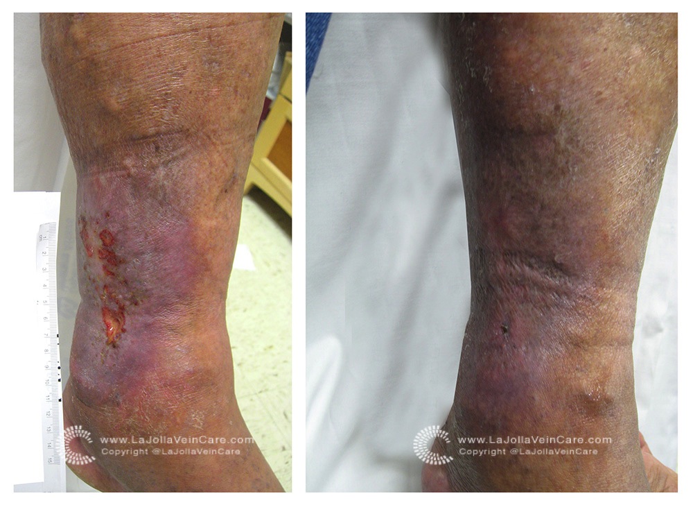
VENOUS LEG ULCER, BEFORE: This is a venous leg ulcer, caused by venous reflux or venous insufficiency. In this picture, you can see varicose veins above the wound.

After

VENOUS LEG ULCER, AFTER: Because this individual had reflux only in the superficial varicose veins, treatment consisted of foam sclerotherapy of the varicosities and compression with unna boot dressing. The wound healed rapidly and note that the varicose veins are gone.

A venous leg ulcer is an open wound on the leg, caused by long-standing vein problems. It is the most severe form of chronic venous insufficiency (CVI). Venous leg ulcers are common, accounting for over 70% of all leg wounds. It affects 1% of Americans and is the seventh leading cause for disability in the United States.

It is usually located around the ankle (on the inner or outer sides). It is caused by underlying venous insufficiency, or venous reflux.  The persistent venous reflux (back flow of blood through faulty valves) causes high pressure within the leg veins.  The high pressure, is then exerting outward on the skin, causing an inflammatory response, eventually causing the skin to break down.  Visible signs of venous disease are varicose veins, ankle swelling, skin discoloration around the ankle and usually precede the leg wound.  Underlying venous insufficiency can be detected by duplex ultrasound imaging (a non-painful study).

The treatment of venous leg ulcers is to correct the underlying problem: the faulty veins. Treatment of the underlying non-functional veins will reduce pressure from venous hypertension, allowing the wound to heal.  There are a number of non-surgical treatment options that can accomplish this including foam sclerotherapy and endovenous ablation, depending on the affected veins. To determine if your leg wound is related to a vein problem or for treatment options, call us for more information.

Can Varicose Veins Cause Leg Ulcers?2021-11-03T23:17:13-07:00

How many veins can be treated before I run out?

2021-11-04T16:49:42-07:00


Before

Before


The veins that can be treated are in the superficial venous system. These veins are located in the subcutaneous tissue and are only responsible for a very small amount of blood return from the legs. All of the diseased superficial veins can be treated because once these veins are closed, the blood is re-routed into the deep veins. The deep system is a more competent system and can accommodate all of the blood from the superficial veins. So, every last problematic superficial vein can be treated with great success.

How many veins can be treated before I run out?2021-11-04T16:49:42-07:00

What If I need my Great Saphenous Vein for Bypass Someday?

2021-11-04T16:18:45-07:00
GSV1

The Great Saphenous Vein is a vein of the superficial venous system that drains blood from the skin and tissues into the deep system. It is most commonly the root of the problem in people with varicose veins. When the great saphenous vein is dysfunctional, removing it is the first step of vein treatment.

A great saphenous vein that needs treatment is not functioning properly. When a vein is diseased, the walls become weak and the vessel can dilate or grow triple its’ normal size. This is not the type of vessel that would be ideal to place in a diseased heart. While the saphenous vein is a common vein that is harvested for bypass grafts, there are other options such as the internal thoracic artery in the chest and the radial artery which is in the arm. With these options, an artery replaces an artery and the removal of the valves inside the veins is not necessary. It is important for the entire circulatory system to be functioning properly, together.

What If I need my Great Saphenous Vein for Bypass Someday?2021-11-04T16:18:45-07:00

Non-surgical Varicose Vein Treatments Mean Quicker Recovery

2021-11-04T14:21:35-07:00
ljvc-slider-4

One of La Jolla Vein Care’s incredible patients returned to surfing just two weeks after varicose vein treatment.

Over the past decade, advances in the treatment of vein conditions have improved the safety, efficacy, comfort, and success of therapy. Newer procedures have replaced old-fashioned vein stripping surgery.  In 2008, a statement by the American College of Phlebology announced that these newer methods, called endovenous procedures are considered the ‘new standard of care,’ replacing vein stripping surgery. Modern vein treatments are performed in the office, without anesthesia, and return to normal activities is almost immediate.  The average procedure time is 60 minutes.   There are several different treatment options depending on the type of veins, the extent of disease, ultrasound findings, and other factors.  Normal activities and regular walking can be resumed immediately after vein treatments, whereas, return to heavy rigorous exercises, such as running and spinning is about two weeks.


Non-surgical Varicose Vein Treatments Mean Quicker Recovery2021-11-04T14:21:35-07:00

28, 4, 2014

Boston Marathon Winner Finishes in Compression Socks

2014-04-28T23:27:28-07:00

The first American man to win the Boston Marathon since 1983 crossed the finish line last week.  Meb Keflezighi, 38 , won the men’s division while wearing CEP compression, a line of Mediven medical compression stockings. Meb has been publicly wearing CEP compression socks for years, including as the US Olympic marathon runner.

images

CEP Compression

Due to the health benefits that come from compression socks, other athletes are also using compression socks to decrease muscle fatigue and improve performance and recovery. It seems as though every runner is wearing compression socks these days.  Compression is not just for athletes. Compression has been used for decades as the treatment for vein disorders. In the field of venous disease, they are the gold standard in conservative management for vein conditions such as varicose veins and venous insufficiency. For those who have jobs that require prolonged periods of standing or sitting, compression can help with occupational leg swelling and fatigue. They are also recommended during air travel to reduce the risk of deep venous thrombosis (DVT).

CEP Compression socks, like those worn by Meb Keflezighi in the Boston Marathon can be found at CompressRx.com.

Boston Marathon Winner Finishes in Compression Socks2014-04-28T23:27:28-07:00

informacion en español

Visita nuestra página en español

https://ljvascular.com/informacion-en-espanol/

Go to Top